A Case of Isolated Squamous Cell Carcinoma of the Orbit
- Affiliations
-
- 1Department of Ophthalmology, Gachon University of Medicine and Science, Incheon, Korea. cmj@gilhospital.com
- KMID: 2214648
- DOI: http://doi.org/10.3341/jkos.2011.52.6.738
Abstract
- PURPOSE
To report a case of isolated squamous cell carcinoma of the orbit.
CASE SUMMARY
A 75-year-old man with over a 50 pack-year history of smoking presented discomfort and visual disturbance of the left eye for several months. His best-corrected visual acuity was 0.3, intraocular pressure was 9 mm Hg, and extraocular movements were normal. Slit-lamp examinations revealed no specific findings in the anterior segment in the left eye. However, retinal exams showed an oval-shaped, juxtapapillary mass-like lesion associated with retinal folding in the left eye. A huge, distorted echoic mass with an initial prominent spike and low-to-medium internal reflectivity with diminishing amplitude was observed on ocular ultrasonography. Enhanced CT revealed a highly-intense, irregular-circumscribed heterogeneous mass (2.0 x 2.0 x 1.5) in the superomedial quadrant of the left eye. Metastatic workups, including bone scan and CT of the head, neck, chest, and abdomen, were unremarkable. One week after the initial visit, the patient experienced pain and reduced visual acuity (light perception) in the left eye. Following the diagnosis, enucleation with tumor resection and hydroxyapatite implantation was performed. Histopathologic examination revealed a moderated-differentiated squamous cell carcinoma invading the sclera. The patient subsequently underwent radiation treatment and no evidence of recurrence was reported 6 months after surgery.
MeSH Terms
Figure
-
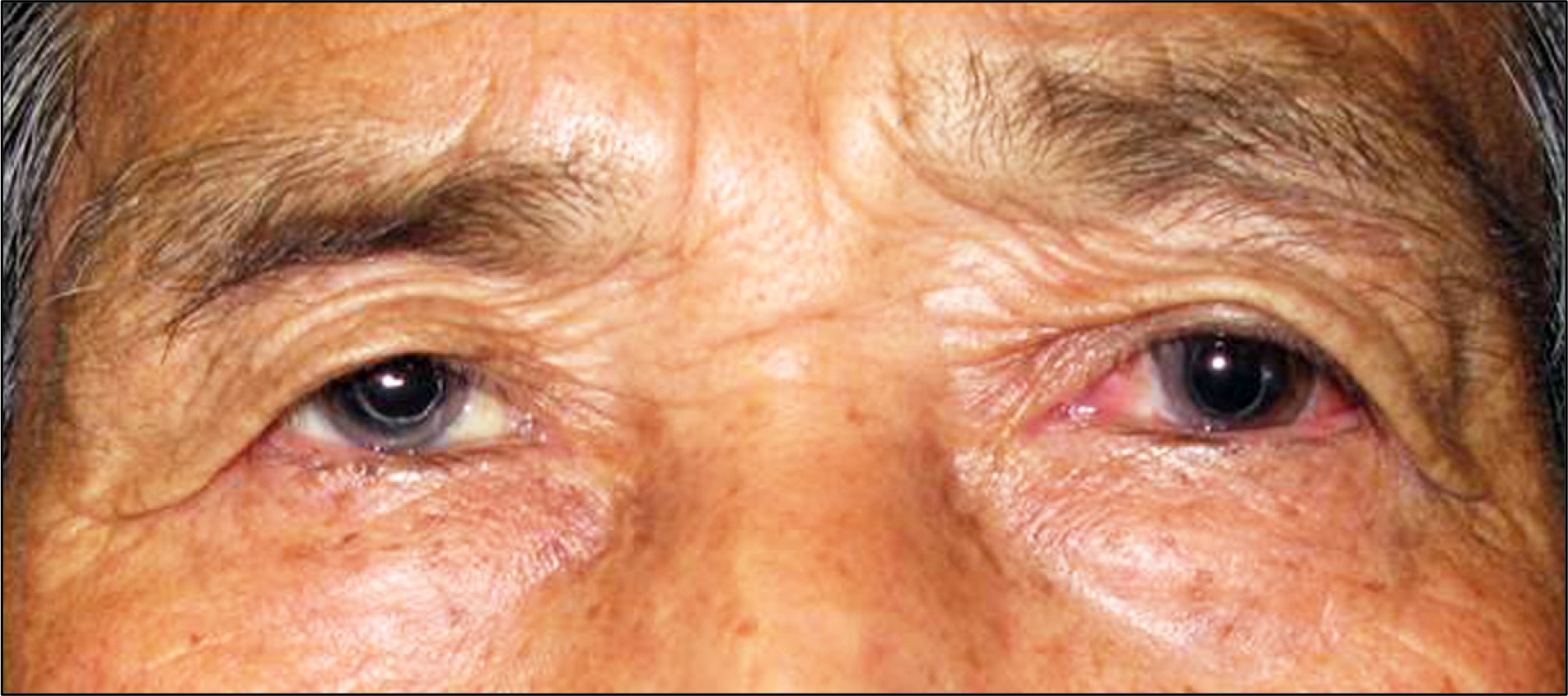
Figure 1. A 75-year-old man without axial proptosis.

Figure 2. An oval shaped, juxtapapillary mass-like lesion with retinal foldings but without dark pigmentations, dragging of retinal vessels, or exudative retinal detachment can be observed.
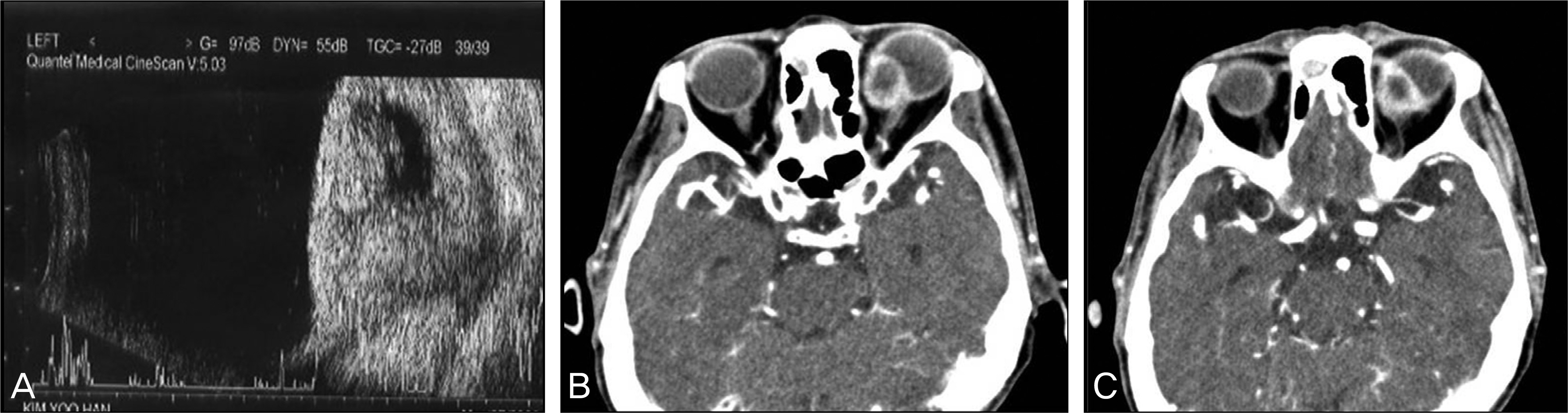
Figure 3. (A). On B-scan, a large mass with distorted echotexture can be seen. A-scan shows an initial prominent spike followed by a low-to-medium internal reflectivity with diminishing amplitude. (B, C) Enhanced axial CT demonstrated a highly-intense, irregularly- circumscribed heterogenous mass (2.0×2.0×1.5 cm sized) in the superomedial quadrant of the left eye.
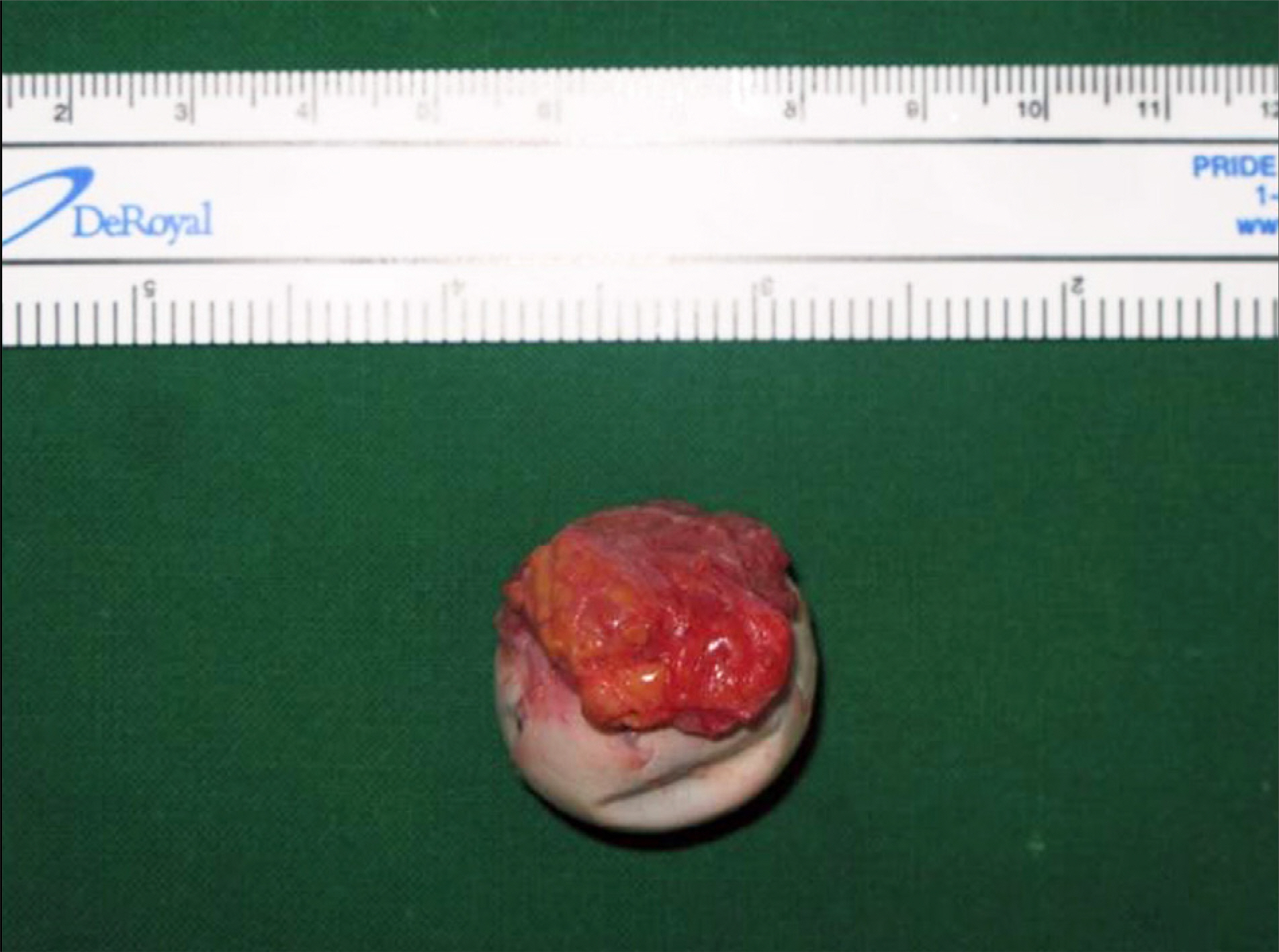
Figure 4. A 2.5×2.5×2.5 cm sized irregular shaped gray- white tumor adjacent to sclera.

Figure 5. (A) Histopathology shows moderately-differentiated squamous cell carcinoma with keratinization (arrow) surrounded by inflammatory cells (asterisk) of lymphocytes, macrophages, neutrophils, eosinophils, and plasma cells (hematoxylin-eosin, ×20). (B) High power view showing numerous pleomorphic cell with hyperchromasia and atypical mitotic figures (hematoxylin-eosin, ×100). (C) Histopathology shows invasion of the sclera (arrow) (hematoxylin-eosin, ×4).

Figure 6. Axial CT shows no evidence of local recurrence 6 months later.
Reference
-
References
1. Ferry AP, Font RL. Carcinoma metastatic to the eye and orbit. I. A clinicopathologic study of 227 cases. Arch Ophthalmol. 1974; 92:276–86.2. Su GW, Patipa M, Font RL. Primary squamous cell carcinoma arising from an epithelium-lined cyst of the lacrimal gland. Ophthal Plast Reconstr Surg. 2005; 21:383–5.
Article3. Holds JB, Anderson RL, Mamalis N, et al. Invasive squamous cell carcinoma arising from asymptomatic choristomatous cysts of the orbit. Two cases and a review of the literature. Ophthalmology. 1993; 100:1244–52.4. McNab AA, Francis IC, Benger R, Crompton JL. Perineural spread of cutaneous squamous cell carcinoma via the orbit. Clinical features and outcome in 21 cases. Ophthalmology. 1997; 104:1457–62.5. Roseman JM. Regression of locally recurrent squamous cell carci-noma of the skin following excision of a metastasis: with review of the literature. J Surg Oncol. 1988; 39:213–4.
Article6. Löffler KU, Witschel H. Orbital squamous cell carcinoma after retinal detachment surgery. Br J Ophthalmol. 1991; 75:568–71.7. Weis E, Shah CP, Lajous M, et al. The association between host susceptibility factors and uveal melanoma: a meta-analysis. Arch Ophthalmol. 2006; 124:54–60.
