Diagnostic Ability of Spectral Domain OCT: Comparision between Preperimetric Glaucoma and Large Physiologic Cupping
- Affiliations
-
- 1Department of Ophthalmology and Inha Vision Science Laboratory, Inha University School of Medicine, Incheon, Korea. nrkim@inha.ac.kr
- KMID: 2214556
- DOI: http://doi.org/10.3341/jkos.2015.56.9.1400
Abstract
- PURPOSE
To assess the distinction ability for differentiating glaucoma patients based on optic disc, retinal nerve fiber layer (RNFL) and ganglion cell-inner plexiform layer (GCIPL) measured using spectral domain optical coherence tomography (SD-OCT). Additionally, the diagnostic ability of these parameters was evaluated by comparing preperimetic glaucoma patients who frequently visited the clinic and normal patients with and without a large physiologic cup/disc (C/D) ratio.
METHODS
Using SD-OCT, the optic disc, RNFL and GCIPL of preperimetic glaucoma patients were compared with normal people with and without a large C/D ratio from March, 2011 to December, 2014 at Department of Ophthalmology, Inha University Hospital. Preperimetic glaucoma was defined using the normal standard automated perimetry for glaucomatous optic nerve changes such as asymmetry of vertical C/D ratio, rim thinning, notching, excavation and RNFL defect.
RESULTS
When comparing preperimetic glaucoma patients to normal people without large disc cupping, the most reliable parameter for optic disc parameters, vertical C/D ratio (0.89), showed more reliable diagnostic ability than the most reliable parameter for retinal nerve fiber, inferior RNFL thickness (0.79) and superonasal and inferonasal GCIPL thickness were the most reliable GCIPL parameters (p = 0.005 and 0.002, respectively). When comparing preperimetic glaucoma patients to normal people having a large physiologic disc cupping, average C/D ratio among optic nerve parameters, inferior RNFL thickness among RNFL thickness parameters and inferior GCIPL thickness among GCIPL parameters showed highly reliable diagnostic abilities. These 3 parameters were not statistically different (all p > 0.05) and had lower distinction ability than reported in previous studies.
CONCLUSIONS
Diagnostic abilities of SD-OCT for distinguishing preperimetric glaucoma from normal people having large physiologic disc cupping were similar for optic disc, RNFL and GCIPL and showed low diagnostic ability than compared to normal people without large disc cupping.
MeSH Terms
Figure
-
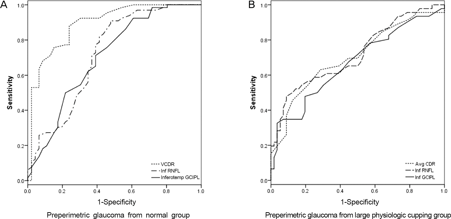
Figure 1. Comparision of receiver operating curves between ONH, RNFL and GCIPL parameters. (A) AUROC values: 0.89 in VCDR, 0.72 in Inf RNFL, 0.70 in GCIPL. The difference between VCDR and Inf RNFL, VCDR and Inferotemp GCIPL were stat-istically significant ( p < 0.001, DeLong method). (B) AUROC values: 0.72 in Inf RNFL, 0.70 in Avg CDR, 0.67 in Inf GCIPL. The difference between Inf RNFL and Avg CDR and Inf GCIPL were not statistically significant ( p > 0.05, DeLong method). ONH = optic nerve head; RNFL = retinal nerve fiber layer; GCIPL = ganglion cell inner plexiform layer; AUROC = area under receiver operating curve; VCDR = vertical cup-disc ratio; Inf RNFL = inferior retinal nerve fiber layer; Inferotemp = in-ferotemporal; Avg CDR = average cup-to-disc ratio; Inf GCIPL = inferior ganglion cell inner plexiform layer.
Reference
-
References
1. Sommer A, Miller NR, Pollack I. . The nerve fiber layer in the diagnosis of glaucoma. Arch Ophthalmol. 1977; 95:2149–56.
Article2. Quigley HA, Addicks EM, Green WR. Optic nerve damage in hu-man glaucoma. III. Quantitative correlation of nerve fiber loss and visual field defect in glaucoma, ischemic neuropathy, papilledema, and toxic neuropathy. Arch Ophthalmol. 1982; 100:135–46.3. Hayreh SS. Pathogenesis of cupping of the optic disc. Br J Ophthalmol. 1974; 58:863–76.
Article4. Quigley HA, Brown AE, Morrison JD, Drance SM. The size and shape of the optic disc in normal human eyes. Arch Ophthalmol. 1990; 108:51–7.
Article5. Carpel EF, Engstrom PF. The normal cup-disk ratio. Am J Ophthalmol. 1981; 91:588–97.
Article6. Armaly MF. Genetic determination of cup/disc ratio of the optic nerve. Arch Ophthalmol. 1967; 78:35–43.7. Armaly MF, Sayegh RE. The cup-disc ratio. The findings of ton-ometry and tonography in the normal eye. Arch Ophthalmol. 1969; 82:191–6.8. Varma R, Tielsch JM, Quigley HA. . Race-, age-, gender-, and refractive error-related differences in the normal optic disc. Arch Ophthalmol. 1994; 112:1068–76.
Article9. Garvin MK, Abràmoff MD, Wu X. . Automated 3-D intra-retinal layer segmentation of macular spectral-domain optical co-herence tomography images. IEEE Trans Med Imaging. 2009; 28:1436–47.
Article10. Bagga H, Feuer WJ, Greenfield DS. Detection of psychophysical and structural injury in eyes with glaucomatous optic neuropathy and normal standard automated perimetry. Arch Ophthalmol. 2006; 124:169–76.
Article11. Jonas JB, Gusek GC, Naumann GO. Optic disc, cup and neuro-retinal rim size, configuration and correlations in normal eyes. Invest Ophthalmol Vis Sci. 1988; 29:1151–8.12. Mwanza JC, Durbin MK, Budenz DL. . Profile and predictors of normal ganglion cell-inner plexiform layer thickness measured with frequency-domain optical coherence tomography. Invest Ophthalmol Vis Sci. 2011; 52:7872–9.
Article13. Kim NR, Lee ES, Seong GJ. . Spectral-domain optical coher-ence tomography for detection of localized retinal nerve fiber layer defects in patients with open-angle glaucoma. Arch Ophthalmol. 2010; 128:1121–8.
Article14. Hanley JA, McNeil BJ. A method of comparing the areas under re-ceiver operating characteristic curves derived from the same cases. Radiology. 1983; 148:839–43.
Article15. Knight OJ, Chang RT, Feuer WJ, Budenz DL. Comparison of reti-nal nerve fiber layer measurements using time domain and spectral domain optical coherent tomography. Ophthalmology. 2009; 116:1271–7.
Article16. Sung KR, Kim DY, Park SB, Kook MS. Comparison of retinal nerve fiber layer thickness measured by Cirrus HD and Stratus op-tical coherence tomography. Ophthalmology. 2009; 116:1264–70. 1270.e1.
Article17. Tan O, Chopra V, Lu AT. . Detection of macular ganglion cell loss in glaucoma by Fourier-domain optical coherence tomography. Ophthalmology. 2009; 116:2305–14.e1-2.
Article18. Na JH, Lee K, Lee JR. . Detection of macular ganglion cell loss in preperimetric glaucoma patients with localized retinal nerve fi-bre defects by spectral-domain optical coherence tomography. Clin Experiment Ophthalmol. 2013; 41:870–80.
Article19. Jeoung JW, Park KH. Comparison of Cirrus OCT and Stratus OCT on the ability to detect localized retinal nerve fiber layer defects in preperimetric glaucoma. Invest Ophthalmol Vis Sci. 2010; 51:938–45.
Article20. Hirashima T, Hangai M, Nukada M. . Frequency-doubling technology and retinal measurements with spectral-domain optical coherence tomography in preperimetric glaucoma. Graefes Arch Clin Exp Ophthalmol. 2013; 251:129–37.
Article21. Rao HL, Kumbar T, Addepalli UK. . Effect of spectrum bias on the diagnostic accuracy of spectral-domain optical coherence to-mography in glaucoma. Invest Ophthalmol Vis Sci. 2012; 53:1058–65.
Article22. Rao HL, Addepalli UK, Chaudhary S. . Ability of different scanning protocols of spectral domain optical coherence tomog-raphy to diagnose preperimetric glaucoma. Invest Ophthalmol Vis Sci. 2013; 54:7252–7.
Article23. Lisboa R, Paranhos A Jr, Weinreb RN. . Comparison of differ-ent spectral domain OCT scanning protocols for diagnosing pre-perimetric glaucoma. Invest Ophthalmol Vis Sci. 2013; 54:3417–25.
Article24. Leite MT, Rao HL, Zangwill LM. . Comparison of the diag-nostic accuracies of the Spectralis, Cirrus, and RTVue optical co-herence tomography devices in glaucoma. Ophthalmology. 2011; 118:1334–9.
Article25. Savini G, Carbonelli M, Barboni P. Retinal nerve fiber layer thick-ness measurement by Fourier-domain optical coherence tomog-raphy: a comparison between cirrus-HD OCT and RTVue in healthy eyes. J Glaucoma. 2010; 19:369–72.
Article26. Buchser NM, Wollstein G, Ishikawa H. . Comparison of retinal nerve fiber layer thickness measurement bias and imprecision across three spectral-domain optical coherence tomography devices. Invest Ophthalmol Vis Sci. 2012; 53:3742–7.
Article
- Full Text Links
-
- Actions
-
Cited
- CITED
-
- Close
- Share
-
- Similar articles
-
- Diagnostic Ability of Swept-Source and Spectral-Domain Optical Coherence Tomography for Glaucoma
- Stratus OCT, SWAP, Matrix FDT in Preperimetric Glaucoma
- Comparison of Diagnostic Ability of 3D and Stratus Optical Coherence Tomography in Early Glaucoma
- Clinical Usefulness of Spectral-Domain Optical Coherence Tomography in Glaucoma and NAION
- The Structure-function Relationships between Two Different Optical Coherence Tomography in Patients with High Myopic Glaucoma
