J Korean Ophthalmol Soc.
2010 Apr;51(4):598-600. 10.3341/jkos.2010.51.4.598.
A Case of Oncocytoma of the Caruncle
- Affiliations
-
- 1Department of Ophthalmology and Visual Science, Catholic University College of Medicine, Seoul, Korea. yswoph@hanmail.net
- KMID: 2213404
- DOI: http://doi.org/10.3341/jkos.2010.51.4.598
Abstract
- PURPOSE
To report a case of oncocytoma of the caruncle.
CASE SUMMARY
A 63-year-old woman visited our clinic due to a mass in her left caruncle that developed five months prior to admission. At the time of her visit, her visual acuity was 0.8 in the right eye and 1.0 in the left eye, and there was a small mass in her left eye. She did not have any pain, and no discharge was apparent. All blood sample results were within the normal range. Also, we did not perform any radiologic studies. We administered systemic antibiotics and excised the mass from the caruncle of her left eye. Her biopsy read 'Oval cells with cytoplasm stuffed with eosinophilic granular material'. These findings are compatible with oncocytoma of the caruncle.
CONCLUSIONS
The oncocytoma of the caruncle requires regular follow-up, which differs from the treatment plans for hemangioma, nevi, and cysts.
Keyword
MeSH Terms
Figure
-
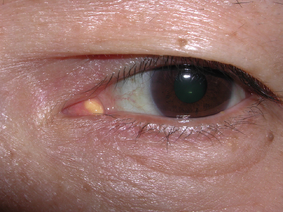
Figure 1. A mass of the caruncle in left eye.

Figure 2. (A) Tumor composed of cells with abundant granular, eosinophilic cytoplasm (Haematoxylin and eosin, magnification ×40). (B) The caruncular stroma displays cystic glandular structures lined by multilayered epithelium (Haematoxylin and eosin, magnification ×200).
Cited by 1 articles
-
A Case of Steatocystoma Simplex and Sebaceous Gland Hyperplasia of the Bilateral Lacrimal Caruncle
Junkyu Chung, Shin-Myeong Choi, Ji Sang Han, Jae-Ho Shin, Tae Gi Kim
J Korean Ophthalmol Soc. 2018;59(9):871-875. doi: 10.3341/jkos.2018.59.9.871.
Reference
-
References
1. Kim KW, Cho YS, Lee KH. Oncocyoma of the minor salivary gland in palate. Chungbuk Medical Journal. 2002; 12:154–60.2. Filho JP, Vianna RN, Coutinho AB, et al. Oncocytoma of the car-uncle; A clinicopathologic case report. Int Ophthalmol. 2004; 25:321–3.
Article3. Archodkis S, Skagias L, Tsakiris A, et al. Oncocytoma of the abdominal gland diagnosed initially by fine-needle aspiration cytology. Diagn Cytopathol. 2009; 37:443–5.4. Calle CA, Castillo IG, Eagle RC. Daza MT. Oncocytoma of the lacrimal gland case report and review of the literature. Orbit. 2006; 25:243–7.5. Pecorella I, Garner A. Ostensible oncocytoma of accessory abdominal glands. Histopathology. 1997; 30:264–70.6. Rennie IG. Oncocytomas(oxyphil adenomas) of the lacrimal caruncle. Br J Ophthalmol. 1980; 64:935–9.7. Ellis GL, Auclair PL. Benign Epithelial Neoplasms.-Tumors of the Salivary Glands. Washington, DC: Armed Forces Institute of Pathology;1996. p. 39–136.8. Biggs SL, Font RL. Oncocytic lesions of the caruncle and other abdominal adnexa. Arch Ophthalmol. 1977; 95:474–8.9. Heathcote JG, Kumalo TG, Willis NR, Mills DM. Oncocytoma of the lacrimal caruncle. Can J Ophthalmol. 1986; 21:178–83.10. Stafford RE, Ray M, Schubert W. Benign oncocytoma of the deep lobe of the parotid gland. J Oral Maxillofac Surg. 1999; 57:346–50.
Article
