Management of Relapsed Inflammatory Choroidal Neovascularization in Punctate Inner Choroidopathy after Bevacizumab
- Affiliations
-
- 1Department of Ophthalmology, Busan Paik Hospital, Inje University College of Medicine, Busan, Korea.
- 2Department of Ophthalmology, Gyeongsang National University School of Medicine, Jinju, Korea. inyoung@gnu.ac.kr
- KMID: 2213255
- DOI: http://doi.org/10.3341/jkos.2016.57.3.513
Abstract
- PURPOSE
To report a rare case of relapsed inflammatory choroidal neovascularization (CNV) in a young female patient after intravitreal bevacizumab (IVB) treatment for subfoveal CNV secondary to punctate inner choroidopathy (PIC).
CASE SUMMARY
A 25-year-old myopic female presented with PIC complicated by subfoveal CNV in the right eye. Her lesion initially responded to three monthly 1.25 mg IVB injections, but the lesion recurred two months after the final injection, and the size of the lesion was larger than that observed before treatment. Further treatment with systemic steroids and IVB resulted in successful anatomic and visual improvement.
CONCLUSIONS
This report presents a rare case of relapsed inflammatory CNV in a young female patient after IVB treatment for subfoveal CNV secondary to PIC. Systemic steroid and IVB were performed after relapse, which successfully improved and maintained vision for longer than 18 months.
MeSH Terms
Figure
-

Figure 1. Fundus photograph, optical coherence tomography, Fluorescein angiography and indocyanine green angiography. (A) A fundus photograph demonstrates multiple, small, yellow, opaque, and round lesions scattered throughout the posterior pole of the right eye. (B) Optical coherence tomography shows the subfoveal choroidal neovascular membrane and subretinal fluid. (C) Fluorescein angiography demonstrates multiple small areas of hyperfluorescence in the right eye. (D) Indocyanine green angiography shows multiple small areas of hypofluorescence in the right eye. (E, F) The patient showed complete resolution of choroidal neovascularization with only a juxtafoveal atrophic scar at one month after the third intravitreal bevacizumab injection.
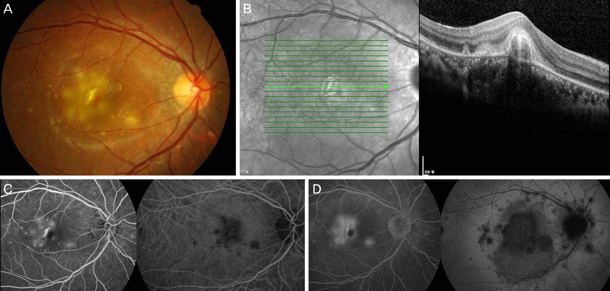
Figure 2. Fundus photograph, optical coherence tomography, Fluorescein angiography and indocyanine green angiography at two months after the final intravitreal bevacizumab injection. Fundus photograph (A) and optical coherence tomography (B) shows a larger number of extended multiple, small, yellow, opaque, and round lesions scattered throughout the posterior pole of the right eye. Fluorescein angiography and indocyanine green angiography (C, D) show more severe choroidal inflammation.
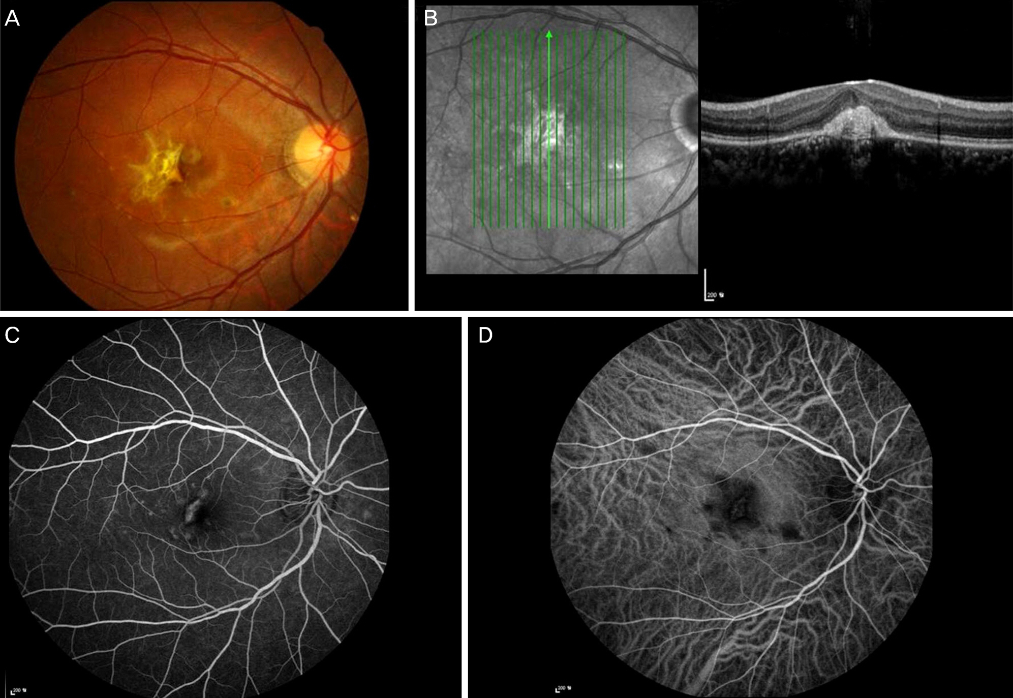
Figure 3. Fundus photograph, optical coherence tomography, Fluorescein angiography and indocyanine green angiography at the final follow-up. Fundus photograph (A), optical coherence tomography (B), fluorescein angiography (C) and indocyanine green angiography (D) show a chorioretinal atrophic scar in the juxtafovea.
Reference
-
References
1. Watzke RC, Packer AJ, Folk JC, et al. Punctate inner choroidopathy. Am J Ophthalmol. 1984; 98:572–84.
Article2. Brown J Jr, Folk JC, Reddy CV, Kimura AE. Visual prognosis of multifocal choroiditis, punctate inner choroidopathy, and the diffuse subretinal fibrosis syndrome. Ophthalmology. 1996; 103:1100–5.
Article3. Jiménez B, Pinilla I, Cristóbal JA, et al. Intravitreal ranibizumab in the treatment of subretinal neovascularization in a case of punctate inner choroidopathy. Arch Soc Esp Oftalmol. 2014; 89:130–2.
Article4. Cho HY, Kim SG, Ham DI, Kang SW. Clinical features of punctate inner choroidopathy in Korea. J Korean Ophthalmol Soc. 2004; 45:2047–54.5. Mansour AM, Arevalo JF, Ziemssen F, et al. Long-term visual outcomes of intravitreal bevacizumab in inflammatory ocular neovascularization. Am J Ophthalmol. 2009; 148:310–16.e2.
Article6. Chan WM, Lai TY, Liu DT, Lam DS. Intravitreal bevacizumab (avastin) for choroidal neovascularization secondary to central serous chorioretinopathy, secondary to punctate inner choroidopathy, or of idiopathic origin. Am J Ophthalmol. 2007; 143:977–83.
Article7. Campos J, Campos A, Beselga D, et al. Punctate inner choroidopathy: a clinical case report. Case Rep Ophthalmol. 2013; 28:155–9.
Article8. Gerstenblith AT, Thorne JE, Sobrin L, et al. Punctate inner choroidopathy: a survey analysis of 77 persons. Ophthalmology. 2007; 114:1201–4.9. Zhang H, Liu ZL, Sun P, Gu F. Intravitreal bevacizumab as primary treatment of choroidal neovascularization secondary to punctate inner choroidopathy: results of a 1-year prospective trial. Retina. 2012; 32:1106–13.10. Arevalo JF, Adan A, Berrocal MH, et al. Intravitreal bevacizumab for inflammatory choroidal neovascularization: results from the Pan-American Collaborative Retina Study Group at 24 months. Retina. 2011; 31:353–63.11. Schouten JS, La Heij EC, Webers CA, et al. A systematic review on the effect of bevacizumab in exudative age-related macular degeneration. Graefes Arch Clin Exp Ophthalmol. 2009; 247:1–11.
Article12. Mansour AM, Arevalo JF, Fardeau C, et al. Three-year visual and anatomic results of administrating intravitreal bevacizumab in inflammatory ocular neovascularization. Can J Ophthalmol. 2012; 47:269–74.
Article13. Mangat SS, Ramasamy B, Prasad S, et al. Resolution of choroidal neovascularization secondary to punctate inner choroidopathy (PIC) with intravitreal anti-VEGF agents: a case series. Semin Ophthalmol. 2011; 26:1–3.
Article14. Cornish KS, Lim LT, Imrie F. Management of inflammatory choroidal neovascularization (CNV) secondary to punctate inner choroidopathy in a young female of childbearing age with intra-vitreal ranibizumab and half-fluence photodynamic therapy (PDT) - a holistic approach. Semin Ophthalmol. 2012; 27:29–32.
Article
- Full Text Links
-
- Actions
-
Cited
- CITED
-
- Close
- Share
-
- Similar articles
-
- Recurrence of Punctate Inner Choroidopathy with an Intravitreal Dexamethasone Implant
- Clinical Features of Punctate Inner Choroidopathy in Korea
- A Case of Punctate Inner Choroidopathy Followed by Multiple Evanescent White Dot Syndrome
- Punctate Inner Choroidopathy
- Choroidal Neovascularization in a Patient with Best Disease
