J Korean Ophthalmol Soc.
2016 Mar;57(3):405-412. 10.3341/jkos.2016.57.3.405.
Comparison of the Visual Outcomes after Cataract Surgery with Implantation of a Bifocal and Trifocal Diffractive Intraocular Lens
- Affiliations
-
- 1Cheil Eye Hospital, Daegu, Korea. eyepark9@naver.com
- KMID: 2213241
- DOI: http://doi.org/10.3341/jkos.2016.57.3.405
Abstract
- PURPOSE
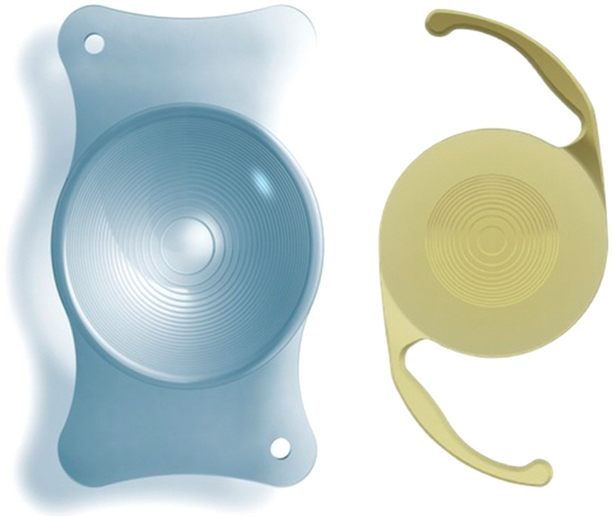
To evaluate and compare visual outcomes and optical quality after implantation of a bifocal (Acrysof ReSTOR® SN6AD1) or trifocal (AT LISA® tri 839MP) diffractive intraocular lens (IOL).
METHODS
Fifty-one eyes of 43 patients undergoing cataract surgery were enrolled and assigned to one of two groups: the trifocal group, comprising 24 eyes implanted with the trifocal diffractive IOL (AT LISA® tri 839MP), and the bifocal group, comprising 27 eyes implanted with the bifocal diffractive IOL (Acrysof ReSTOR® SN6AD1). Visual acuity (distant, intermediate, and near vision) and refractive postoperative outcomes were evaluated at one and three months postoperatively. Measurements of optical quality (using OQAS II®), contrast sensitivity (using CGT-2000®), automated visual field examination, and evaluation of defocus curve were performed three months postoperatively.
RESULTS
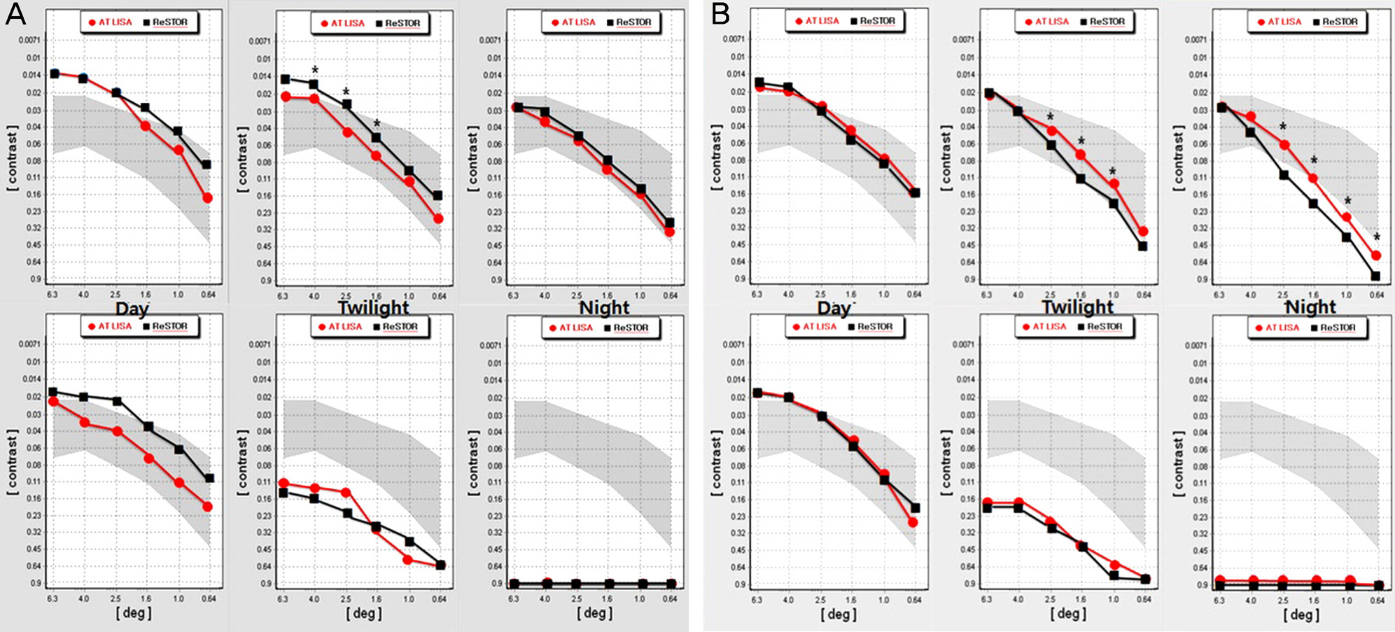
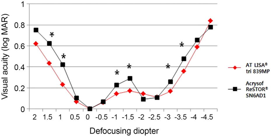
No statistically significant differences between the two groups were found in three-month postoperative distant and near (40 cm) visual acuities and optical quality. However, intermediate (63 cm, 80 cm, and 100 cm) visual acuities were significantly better in the trifocal group. Distant contrast sensitivity (5 m) under mesopic conditions was significantly better with the bifocal lens, whereas near contrast sensitivity (30 cm) under mesopic and scotopic conditions was significantly better with trifocal lens. There was no statistical difference between the groups under photopic conditions. In the defocus curve, the visual acuity was significantly better at intermediate distance in the trifocal group.
CONCLUSIONS
Trifocal diffractive IOLs provide significantly better intermediate vision than bifocal IOLs, with equivalent postoperative levels of distant and near vision and ocular optical quality. Further, they provide better near contrast sensitivity under scotopic condition compared to diffractive bifocal IOLs.
Keyword
Figure
Reference
-
References
1. Davison JA, Simpson MJ. History and development of the apodized diffractive intraocular lens. J Cataract Refract Surg. 2006; 32:849–58.
Article2. Alfonso JF, Fernández-Vega L, Baamonde MB, Montés-Micó R. Prospective visual evaluation of apodized diffractive intraocular lenses. J Cataract Refract Surg. 2007; 33:1235–43.
Article3. de Vries NE, Webers CA, Montés-Micó R, et al. Long-term follow-up of a multifocal apodized diffractive intraocular lens after cataract surgery. J Cataract Refract Surg. 2008; 34:1476–82.
Article4. Park YL, Hwang GY, Joo CK. Comparative study of clinical outcomes between 2 types of multifocal aspheric intraocular lenses. J Korean Ophthalmol Soc. 2013; 54:1199–207.
Article5. Blaylock JF, Si Z, Vickers C. Visual and refractive status at different focal distances after implantation of the ReSTOR multifocal intraocular lens. J Cataract Refract Surg. 2006; 32:1464–73.
Article6. Alfonso JF, Fernández-Vega L, Puchades C, Montés-Micó R. Intermediate visual function with different multifocal intraocular lens models. J Cataract Refract Surg. 2010; 36:733–9.
Article7. Voskresenskaya A, Pozdeyeva N, Pashtaev N, et al. Initial results of trifocal diffractive IOL implantation. Graefes Arch Clin Exp Ophthalmol. 2010; 248:1299–306.
Article8. Gatinel D, Pagnoulle C, Houbrechts Y, Gobin L. Design and qualification of a diffractive trifocal optical profile for intraocular lenses. J Cataract Refract Surg. 2011; 37:2060–7.
Article9. Cochener B, Vryghem J, Rozot P, et al. Visual and refractive outcomes after implantation of a fully diffractive trifocal lens. Clin Ophthalmol. 2012; 6:1421–7.
Article10. Sheppard AL, Shah S, Bhatt U, et al. Visual outcomes and subjective experience after bilateral implantation of a new diffractive trifocal intraocular lens. J Cataract Refract Surg. 2013; 39:343–9.
Article11. Alfonso JF, Puchades C, Fernández-Vega L, et al. Contrast sensitivity comparison between AcrySof ReSTOR and Acri. LISA aspheric intraocular lenses. J Refract Surg. 2010; 26:471–7.12. Alfonso JF, Fernández-Vega L, Blázquez JI, Montés-Micó R. Visual function comparison of 2 aspheric multifocal intraocular lenses. J Cataract Refract Surg. 2012; 38:242–8.
Article13. Kim SM, Kim CH, Chung ES, Chung TY. Visual outcome and patient satisfaction after implantation of multifocal IOLs: three-month follow-up results. J Korean Ophthalmol Soc. 2012; 53:230–7.
Article14. Kim JH, Kim EJ, Kim YI, et al. Comparison of clinical outcomes between diffractive and refractive multifocal intraocular lens with same near added. J Korean Ophthalmol Soc. 2015; 56:875–84.
Article15. Kretz FT, Gerl M, Gerl R, et al. Clinical evaluation of a new pupil independent diffractive multifocal intraocular lens with a +2. 75 D near addition: a European multicentre study. Br J Ophthalmol. 2015; May. pii. bjophthalmol-2015-306811. [Epub ahead of print].16. Marques EF, Ferreira TB. Comparison of visual outcomes of 2 diffractive trifocal intraocular lenses. J Cataract Refract Surg. 2015; 41:354–63.
Article17. Mojzis P, Kukuckova L, Majerova K, et al. Comparative analysis of the visual performance after cataract surgery with implantation of a bifocal or trifocal diffractive IOL. J Refract Surg. 2014; 30:666–72.
Article18. Cabot F, Saad A, McAlinden C, et al. Objective assessment of crystalline lens opacity level by measuring ocular light scattering with a double-pass system. Am J Ophthalmol. 2013; 155:629–35. 635.e1-2.
Article19. Lee K, Ahn JM, Kim EK, Kim TI. Comparison of optical quality parameters and ocular aberrations after wavefront-guided laser in-situ keratomileusis versus wavefront-guided laser epithelial keratomileusis for myopia. Graefes Arch Clin Exp Ophthalmol. 2013; 251:2163–9.
Article20. Saad A, Saab M, Gatinel D. Repeatability of measurements with a double-pass system. J Cataract Refract Surg. 2010; 36:28–33.
Article21. Aychoua N, Junoy Montolio FG, Jansonius NM. Influence of multifocal intraocular lenses on standard automated perimetry test results. JAMA Ophthalmol. 2013; 131:481–5.
Article22. Farid M, Chak G, Garg S, Steinert RF. Reduction in mean deviation values in automated perimetry in eyes with multifocal compared to monofocal intraocular lens implants. Am J Ophthalmol. 2014; 158:227–31.e1.
Article23. Lee JM, Seo KY, Kim EK. Comparison of optical aberrations and contrast sensitivity between monofocal and multifocal intraocular lens. J Korean Ophthalmol Soc. 2002; 43:1882–6.24. Yoon JU, Chung JL, Hong JP, et al. Comparison of wavefront analysis and visual function between monofocal and multifocal aspheric intraocular lenses. J Korean Ophthalmol Soc. 2009; 50:195–201.
Article25. Kamiya K, Hayashi K, Shimizu K, et al. Multifocal intraocular lens explantation: a case series of 50 eyes. Am J Ophthalmol. 2014; 158:215–20.e1.26. Alfonso JF, Fernández-Vega L, Baamonde MB, Montés-Micó R. Correlation of pupil size with visual acuity and contrast sensitivity after implantation of an apodized diffractive intraocular lens. J Cataract Refract Surg. 2007; 33:430–8.
Article
- Full Text Links
- Actions
-
Cited
- CITED
-
- Close
- Share
- Similar articles
-
- Trifocal versus Bifocal Diffractive Intraocular Lens Implantation after Cataract Surgery or Refractive Lens Exchange: a Meta-analysis
- Comparison of Reading Speed after Bilateral Bifocal and Trifocal Intraocular Lens Implantation
- Effects of Bifocal versus Trifocal Diffractive Intraocular Lens Implantation on Visual Quality after Cataract Surgery
- Clinical Outcomes of Patients Fitted with Bifocal and Trifocal Diffractive Intraocular Lenses
- Clinical Outcomes of Diffractive Aspheric Trifocal Intraocular Lens Implantation


