Multiple Retinal Arterial Occlusion and Retinal Vasculitis Associated With IgA Nephropathy
- Affiliations
-
- 1Department of Ophthalmology, Gyeong Sang National University, College of Medicine, Jinju, Korea. parkjm@gnu.ac.kr
- 2Gyeong Sang Institute of Health Science, Gyeongsang National University, Jinju, Korea.
- KMID: 2213004
- DOI: http://doi.org/10.3341/jkos.2009.50.12.1887
Abstract
- PURPOSE
To report a case of occlusion of multiple retinal arteries associated with retinal vasculitis-associated IgA nephropathy.
Case summary
A 37-year-old diagnosed with immunoglobulin A(IgA) nephropathy by renal biopsy four months prior was referred to an ophthalmologist complaining of visual impairment in her right eye. On physical examination, the patient's visual acuity was hand-movements in the right eye and 1.0 in the left eye. She had relative afferent papillary defect in the right eye with cells in the anterior chamber. She was diagnosed with occlusion of multiple retinal arteries and retinal vasculitis on fundus examination and fluorescein angiogram. There were no abnormal findings on routine hematologic tests including thrombophilia studies, carotid artery, or cardiovascular examination. Antinuclear antibody, rheumatoid factor, and antiphospholipid antibody were negative.
CONCLUSIONS
The patient was diagnosed with retinal artery occlusion combined with retinal vasculitis associated with IgA nephropathy.
MeSH Terms
-
Adult
Anterior Chamber
Antibodies, Antinuclear
Antibodies, Antiphospholipid
Biopsy
Carotid Arteries
Eye
Fluorescein
Glomerulonephritis, IGA
Hematologic Tests
Humans
Immunoglobulin A
Immunoglobulins
Physical Examination
Retinal Artery
Retinal Artery Occlusion
Retinal Vasculitis
Retinaldehyde
Rheumatoid Factor
Thrombophilia
Vision Disorders
Visual Acuity
Antibodies, Antinuclear
Antibodies, Antiphospholipid
Fluorescein
Immunoglobulin A
Immunoglobulins
Retinaldehyde
Rheumatoid Factor
Figure
-
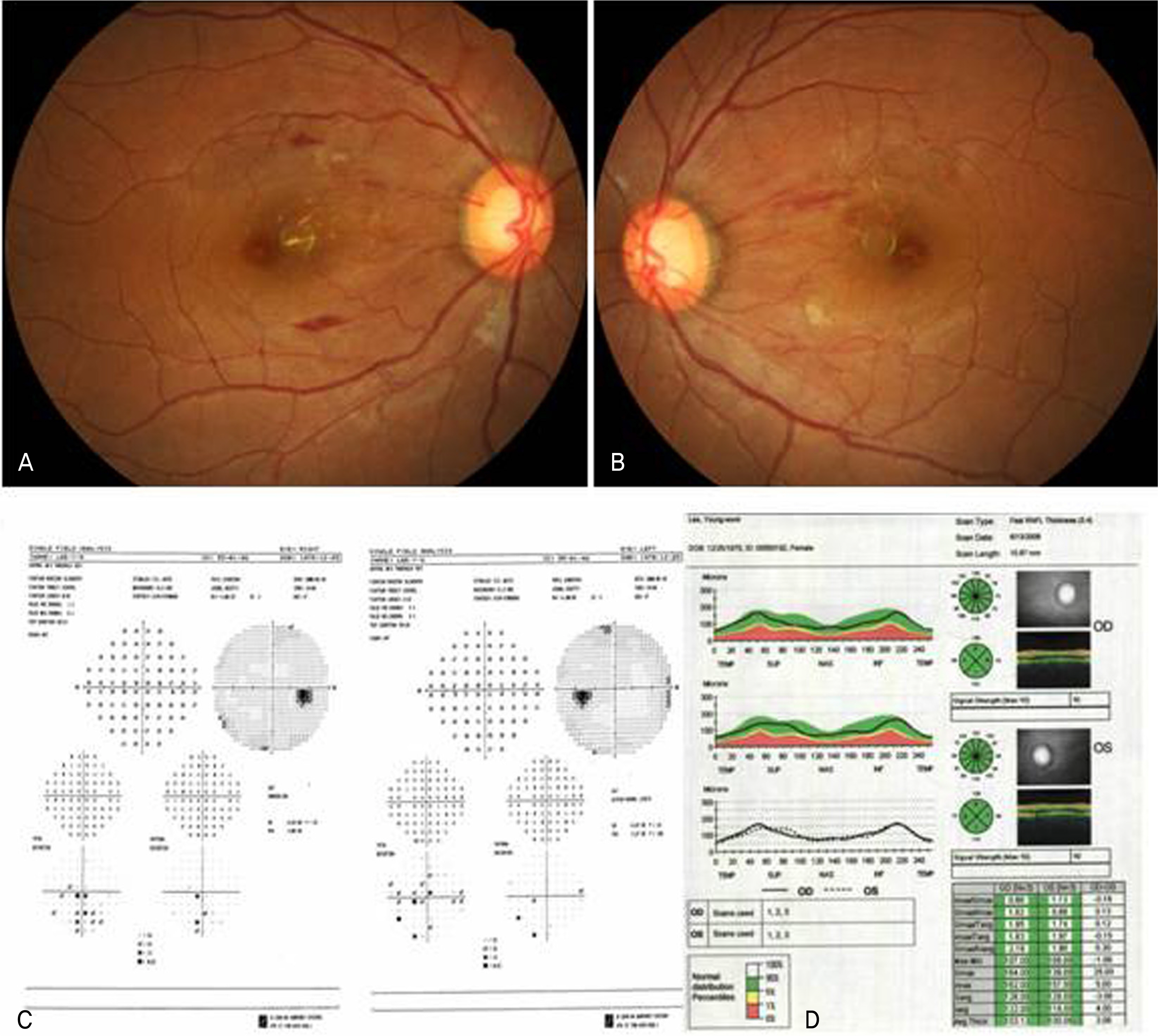
Figure 1. Fundus photograph of both eyes at the first examination. Prominent arteriolar narrowing and multiple retinal hemorrhages and cotton-wool spots can be seen in the retina (A, B). Visual fields were within normal limit (C). RNFL thickness in glaucoma OCT is normal in both eye (D).

Figure 2. (A) Immunofluorescein examination of kidney biopsy shows accumulation of IgA in mesangeal matrix. (B) Electromicroscopic examination shows electron-dense deposit in the mesangium and patchy area of epithelial foot process effacement.
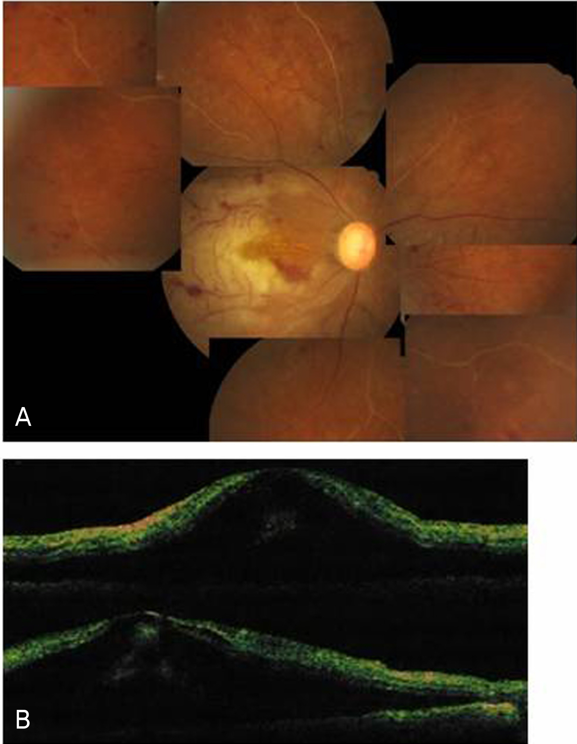
Figure 3. Four months after the intial examination, fundus photograph of the eye shows multiple retinal hemorrhages, obstruction of multiple retinal arteries and ischemic changes of the retina (A). OCT of right eye shows submacular exudation (B).
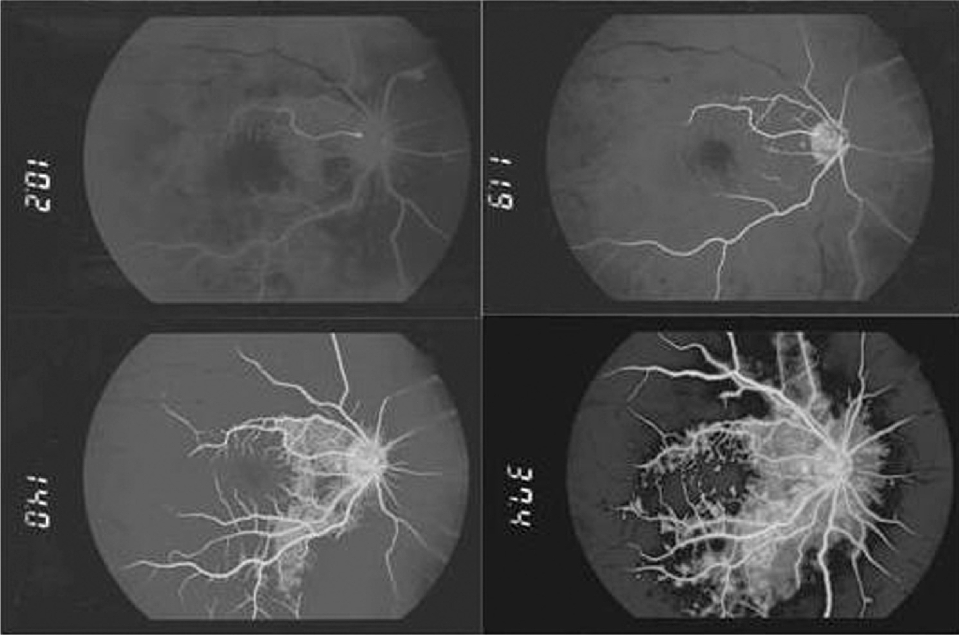
Figure 4. Fluorescein angiogram of the right fundus shows marked delay of filling in the early arterial phase and arteriovenous transit time at the macular and superotemporal area, and severe perivascular fluorescent leakage.
Reference
-
References
1. Fauci AS, Braunwald E, Kasper DL, et al. Harrison's principles of internal medicine. 17th ed.2. New York: McGrow-Hill medical;2008. p. 1788–9.2. Sinniah R. IgA mesangial nephropathy: Berger's disease. Am J Nephrol. 1985; 5:73–83.
Article3. André C, Berthoux FC, André F, et al. Prevalence of IgA2 deposit in IgA nephropathies: A clue to their pathogenesis. N Eng J Med. 1980; 303:1343–6.4. Møller-Jensen J, Marthinsen L, Linné T. Anterior Uveitis in IgA Nephropathy. Am J Ophthalmol. 1989; 108:604–5.
Article5. Izzedine H, Bodaghi B, Launay-Vacher V, Deray G. Oculorenal manifestations in systemic autoimmune disease. Am J Kidney dis. 2004; 43:209–22.6. Nomoto Y, Sakai H, Endoh M, Tomino Y. Scleritis and IgA Nephropathy. Arch Intern Med. 1980; 140:783–5.
Article7. Yamabe H, Ozawa K, Fukushi K. IgA nephropathy and henoch-schonlein purpura nephritis with anterior uveitis. Nephron. 1988; 50:368–70.8. Hurault de Ligny B, Sirbat D, Bene MC, et al. Scleritis associated with glomerulonephritis. Nephron. 1983; 35:207.
Article9. Kaplan HJ, Waldrep JC. Immunologic insights into uveitis and retinitis: the immunoregulatory circuit. Ophthalmology. 1984; 91:655–65.10. Pavlin CJ, Easterbrook M, Harasiewicz K, Foster FS. An ultrasound biomicroscopic analysis of angle-closure glaucoma secondary to ciliochoroidal effusion in IgA nephropathy. Am J Ophthalmol. 1993; 116:341–5.
Article11. Wolfensberger TJ, Piguer B, Gregor ZJ, Bird AC. Retinal vasculopathy associated with Berger's IgA Nephropathy. Klin Monatsbl Augenheilkd. 2000; 216:334–8.12. Kim KJ, Kwak HW. A case of Choroiditis Associated with IgA Nephropathy. J Korean Ophthalmol Soc. 1987; 28:1095–9.13. Kim SK, Kwon SK. Prepapillary vascular loops associated with branch retinal artery occlusion and vitreous hemorrhage. J Korean Ophthalmol Soc. 2007; 48:1001–6.
- Full Text Links
-
- Actions
-
Cited
- CITED
-
- Close
- Share
-
- Similar articles
-
- Prepapillary Vascular Loops associated with Branch Retinal Artery Occlusion and Vitreous Hemorrhage
- Eales Disease Accompanied with Branch Retinal Vein Occlusion
- Retinal Arteriolar Changes in a Patient with Branch Retinal Vein Occlusion
- A Clinical Study of 36 Cases of Retinal Artery Occlusion
- A Case of Tuberculosis-related Retinal Vasculitis
