J Korean Ophthalmol Soc.
2016 May;57(5):752-756. 10.3341/jkos.2016.57.5.752.
Clinical Outcome of Ciliary Sulcus-Implanted Single-Piece Foldable Acrylic Intraocular Lens
- Affiliations
-
- 1Department of Ophthalmology, Yonsei University Wonju College of Medicine, Wonju, Korea. eyedockim@yonsei.ac.kr
- KMID: 2212717
- DOI: http://doi.org/10.3341/jkos.2016.57.5.752
Abstract
- PURPOSE
To evaluate complications and refractive outcomes of implantation of a single-piece acrylic intraocular lens (SPA-IOL) in the ciliary sulcus during phacoemulsification complicated with posterior capsule tear (PCT).
METHODS
This retrospective study included patients who visited our hospital from 2014 January to 2015 June with implantation of a SPA-IOL (RAYNER 920H Superflex) in the ciliary sulcus during phacoemulsification complicated with PCT. Patients had their IOL power reduced by 1 diopter (D) from that calculated for in-the-bag implantation. At 3 months after operation, best corrected visual acuity (BCVA), uncorrected visual acuity (UCVA) and a refraction test were performed.
RESULTS
PCT requiring implantation of IOL in the ciliary sulcus occurred in 17 eyes. Postoperative complications included corneal edema (3 eyes), anterior segment inflammation (1 eye), intraocular pressure elevations (3 eyes). However, improvements resulting from proper management and no significant IOL decentration were observed. At 3 months after operation, the mean spherical equivalent was -0.79 ± 0.39 D (-0.25 to -1.5 D), the mean UCVA was 0.77 ± 0.22 (0.4 to 1.0), and the mean BCVA was 0.94 ± 0.08 (0.8 to 1.0).
CONCLUSIONS
Sulcus implantation of a SPA-IOL (RAYNER 920H Superflex) has no clinically significant complication, and the mean spherical equivalent after 3 months with a power reduction of 1.0 D was -0.79 ± 0.39 D (-0.25 to -1.5 D).
MeSH Terms
Figure
-
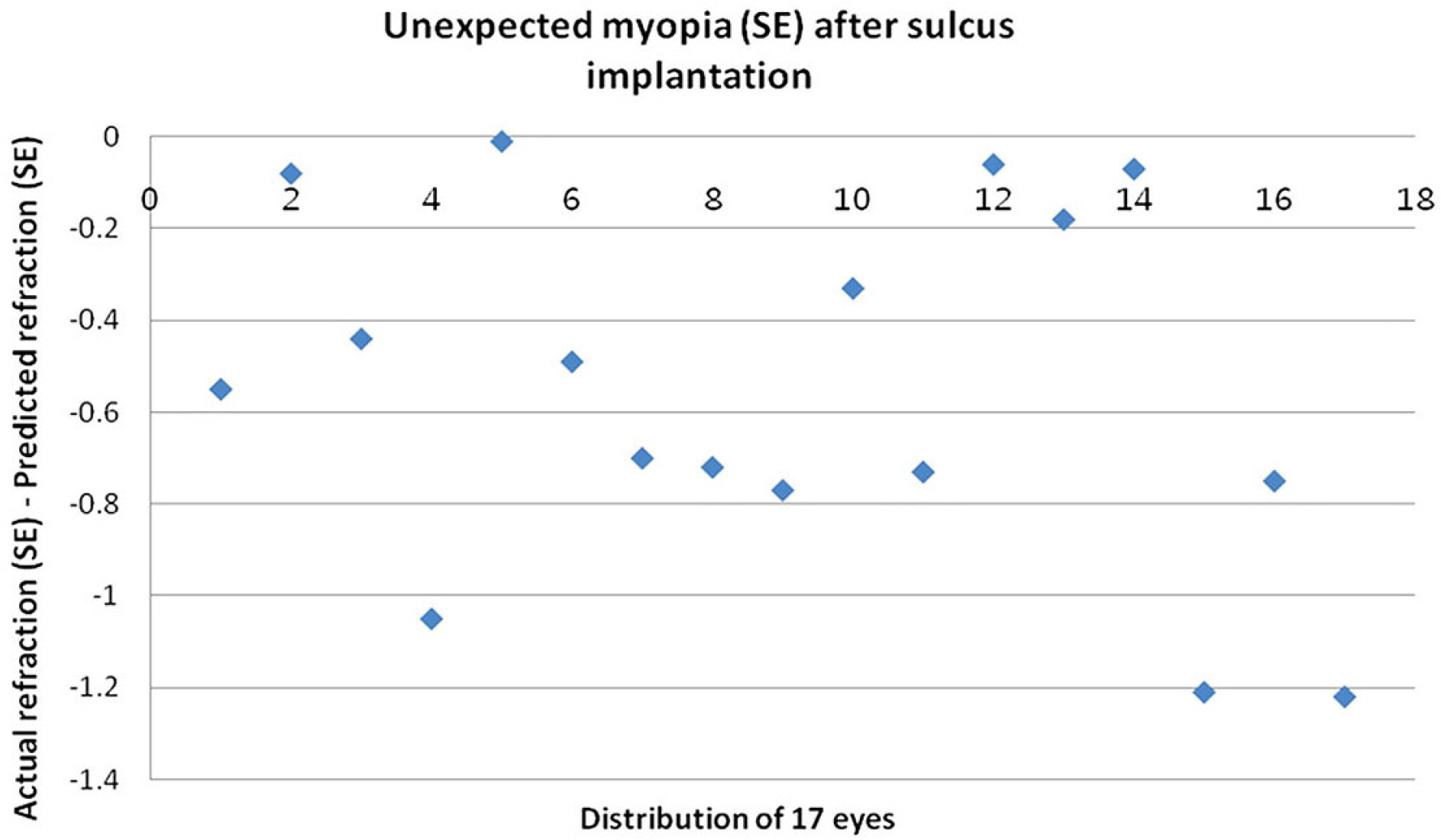
Figure 1. Unexpected myopia. Diagram demonstrating the unexpected myopia (SE) after sulcus implantation of a single-piece foldable acrylic intraocular lens with 1 D less power than that calculated for in-the-bag fixation. SE = spherical equivalent.
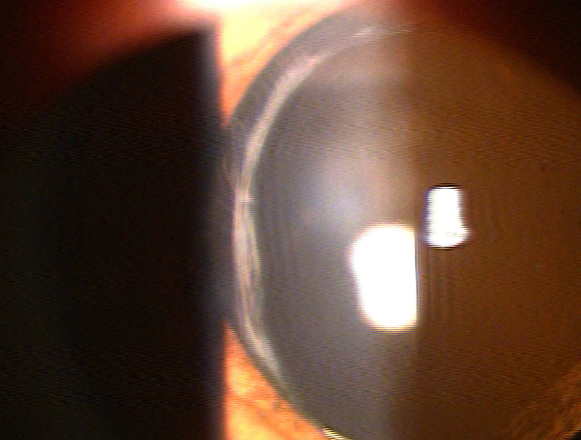
Figure 2. Slit-lamp examination. Photograph showing a centered ciliary sulcus intraocular lens.
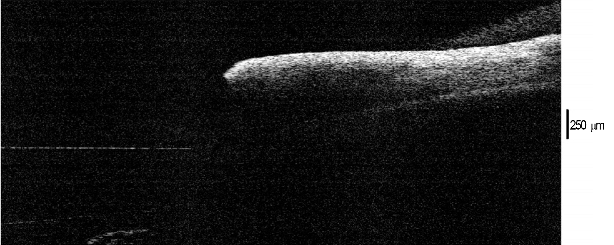
Figure 3. Optical coherence tomography shows no iris-intraocular lens (IOL) contact.
Reference
-
References
1. Chang DF. Single versus three piece acrylic IOLs. Br J Ophthalmol. 2004; 88:727–8.
Article2. Schulze S. Bertelmann T. Sekundo W. Implantation of intraocular lenses in the ciliary sulcus. Ophthalmologe. 2014; 111:305–9.3. Chang DF. Masket S. Miller KM, et al. Complications of sulcus placement of single-piece acrylic intraocular lenses: recommendations for backup IOL implantation following posterior capsule rupture. J Cataract Refract Surg. 2009; 35:1445–58.4. Mamalis N. Sulcus placement of single-piece acrylic intraocular lenses. J Cataract Refract Surg. 2009; 35:1327–8.
Article5. Dubey R. Birchall W. Grigg J. Improved refractive outcome for ciliary sulcus-implanted intraocular lenses. Ophthalmology. 2012; 119:261–5.
Article6. Bar-Sela SM. Fleissig E. Intermediate term follow-up after a single-piece-acrylic intraocular lens implantation in the ciliary sulcus- a cross-sectional study. BMC Ophthalmol. 2013; 13:76.
Article7. Hong Y. Sun YX. Qi H, et al. Pigment dispersion glaucoma induced by the chafing effect of intraocular lens haptics in Asian eyes. Curr Eye Res. 2013; 38:358–62.
Article8. Renieri G. Herzog D. Niemann S, et al. Sulcus implantation of a single-piece foldable acrylic intraocular lens after posterior capsular rupture in cataract surgery. Eur J Ophthalmol. 2012; 22:950–5.
Article9. Ionides A. Minassian D. Tuft S. Visual outcome following posterior capsule rupture during cataract surgery. Br J Ophthalmol. 2001; 85:222–4.
Article10. Zarei-Ghanavati S. Gharaii H. Zarei-Ghanavati M. Simple method to evaluate adequacy of capsule for foldable intraocular lens implantation in the sulcus. J Cataract Refract Surg. 2009; 35:222–5.
Article11. Taskapili M. Engin G. Kaya G, et al. Single-piece foldable acrylic intraocular lens implantation in the sulcus in eyes with posterior capsule tear during phacoemulsification. J Cataract Refract Surg. 2005; 31:1593–7.
Article12. Kirk KR. Werner L. Jaber R, et al. Pathologic assessment of complications with asymmetric or sulcus fixation of square-edged hydrophobic acrylic intraocular lenses. Ophthalmology. 2012; 119:907–13.
Article13. Uy HS. Chan PS. Pigment release and secondary glaucoma after implantation of single-piece acrylic intraocular lenses in the ciliary sulcus. Am J Ophthalmol. 2006; 142:330–2.
Article14. LeBoyer RM. Werner L. Snyder ME, et al. Acute haptic-induced ciliary sulcus irritation associated with single-piece AcrySof intraocular lenses. J Cataract Refract Surg. 2005; 31:1421–7.
Article15. Micheli T. Cheung LM. Sharma S, et al. Acute haptic-induced pigmentary glaucoma with an AcrySof intraocular lens. J Cataract Refract Surg. 2002; 28:1869–72.
Article16. Kohnen T. Kook D. Solving intraocular lens-related pigment dispersion syndrome with repositioning of primary sulcus implanted single-piece IOL in the capsular bag. J Cataract Refract Surg. 2009; 35:1459–63.
Article17. Birner B. Tourtas T. Wessel JM, et al. Pigment dispersion syndrome and pigmentary glaucoma. Morphometric analysis of the anterior chamber segment with SL-OCT. Ophthalmologe. 2014; 111:638–43.18. Niyadurupola N. Broadway DC. Pigment dispersion syndrome and pigmentary glaucoma-a major review. Clin Experiment Ophthalmol. 2008; 36:868–82.19. Wintle R. Austin M. Pigment dispersion with elevated intraocular pressure after AcrySof intraocular lens implantation in the ciliary sulcus. J Cataract Refract Surg. 2001; 27:642–4.
Article
- Full Text Links
-
- Actions
-
Cited
- CITED
-
- Close
- Share
-
- Similar articles
-
- Suture Fixation Technique for a Single-piece Foldable Closed-loop Intraocular Lens
- Intraocular pressure after extracapsular cataract extraction with implantation of posterior chamber lenses.
- Modified Surgical Technique for Transscleral Fixation of a Single-Piece Acrylic Intraocular Lens in the Absence of Capsular Support
- Scleral Fixation of Foldable Posterior Chamber Intraocular Lenses
- Clinical Outcomes of Aspheric 1-Piece (Tecnis(R) ZCB00) and 3-Piece (Tecnis(R) ZA9003) Aspheric Intraocular Lens for 12 Months
