J Korean Ophthalmol Soc.
2009 Aug;50(8):1266-1269. 10.3341/jkos.2009.50.8.1266.
A Case of Multiple Myeloma Presented With Bilateral Corneal Crystalline Deposition
- Affiliations
-
- 1Department of Ophthalmology, Maryknoll Hospital, Busan, Korea. wansookim@yahoo.com
- KMID: 2212549
- DOI: http://doi.org/10.3341/jkos.2009.50.8.1266
Abstract
- PURPOSE
Multiple myeloma is characterized by the neoplastic proliferation of a single clone of plasma cells. Multiple myeloma rarely involves the eyeball or the orbital tissues. We report a case of multiple myeloma that presented with corneal crystalline depositions in a patient complaining of decreased vision and irritation of both eyes without any systemic symptoms. CASE SUMMARY: A 63-year-old woman complained of decreased vision and irritation of both eyes that had started suddenly 20 days before. Uncorrected visual acuity was 0.2 in the right eye and 0.3 in the left eye. Best-corrected visual acuity (BCVA) was 0.9 in the right eye and 1.0 in the left eye. Slit lamp examination showed gray-white crystalline depositions on the epithelium, stroma and Descemet membrane of the cornea extensively, except for the limbus. There were no specific findings on intraocular pressure measurement and fundus examination. The patient did not complain of any systemic symptoms. Multiple myeloma was diagnosed by blood examination and bone marrow biopsy. Two months later, the BCVA decreased to 0.3 in both eyes and artificial tears were prescribed to relieve irritation. CONCLUSIONS: When irritation and decreased vision occur abruptly even without systemic symptoms, there is a possibility of multiple myeloma. Systemic evaluations are needed in these cases.
MeSH Terms
Figure
-

Figure 1. Slit lamp examination showed (A) numerous, delicate, scintillating gray-white crystalline deposits of various size on (B) the epithelium, stroma and Descemet membrane of the cornea extensively except the limbus.
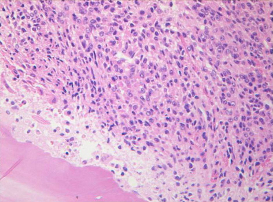
Figure 2. Bone marrow biopsy revealed marrow portion was replaced by immature plasma cells and mature plasma cells with an eccentric nucleus composed of coarsely clumped chromatin and densely basophilic cytoplasm.
Reference
-
References
1. Fauci AS, Braunwald E, Kasper DL, et al. Harrison's principle's of internal medicine. 17th ed.New York: Macgraw-Hill;2008. p. 701–7.2. Knapp AJ, Gartner S, Henkind P, et al. Multiple myeloma and its ocular manifestations. Surv Ophthalmol. 1987; 31:343–51.
Article3. Nakatukasa M, Sotozono C, Taniloka K, et al. Diagnosis of multiple myeloma in a patient with atypical corneal findings. Cornea. 2008; 27:249–51.4. Burki E. Corneal changes in a case of multiple myeloma. Ophthalmologica. 1958; 135:565–72.5. Aronson SB, Shaw R. Corneal crystal in multiple myeloma. Arch Ophthalmol. 1959; 61:541–6.6. Auran JD, Donn A, Hyman GA. Multiple myeloma presenting as vortex crystalline keratopathy and complicated by endocapsular hematoma. Cornea. 1992; 11:584–5.
Article7. Lym CR, Keak NH, Huh W. Central retinal vein occlusion in multiple myeloma associated with hyperviscoity syndrome. J Korean Ophthalmol Soc. 1996; 37:1371–5.8. Shin SW, Shin HH. A case of orbital involvement in multiple myeloma. J Korean Ophthalmol Soc. 1994; 35:474–9.9. Lee MJ, Choung HK, Khwang SI, Yang HJ. Multiple myeloma presented with unilateral ptosis. J Korean Ophthalmol Soc. 2005; 46:1073–8.10. Kim HC, Won IG. A case of orbital involvement in multiple myeloma. J Korean Ophtalmol Soc. 1986; 27:405–10.11. Han HJ, Ma KH. Eye symptoms and signs in multiple myeloma. J Korean Ophthalmol Soc. 1980; 21:627–30.12. Francois J, Rabaey M. Corneal dystrophy and paraproteinemia. Am J Ophthalmol. 1961; 52:895–901.
Article13. Pinkerton RM, Robertson DM. Corneal and conjunctival changes in dysproteinemia. Invest Ophthalmol. 1969; 8:357–64.14. Klintworth GK, Bredenhoeft SJ, Reed JW. Analysis of corneal crystalline deposits in benign monoclonal gammopathy. Am J Ophthalmol. 1978; 86:303–13.15. Silkiss RZ, Pomerleau D, Sorenson A, et al. Corneal cupremia in multiple myeloma: a clinicopathologic correlation. Arch Ophthalmol. 2008; 126:1005–6.
Article16. Lüllmann H, Lüllmann-Rauch R, Wassermann O. Lipidosis induced by amphiphilic cationic drugs. Biochem Pharmacol. 1978; 27:1103–8.
Article17. Rodrigues MM, Krachmer JH, Miller SD, Newsome DA. Poste-rior corneal crystalline deposits in benign monoclonal gammopathy. Arch Ophthalmol. 1979; 97:124–8.
Article18. Beebe WE, Webster RG Jr, Spencer WB. Atypical corneal manifestations of multiple myeloma. Aclinical, histopathologic, and immunohistochemical report. Cornea. 1989; 8:274–80.
