J Korean Ophthalmol Soc.
2009 Jul;50(7):1044-1049. 10.3341/jkos.2009.50.7.1044.
The Clinical Feature of Epiretinal Membrane After Retinal Detachment Surgery
- Affiliations
-
- 1Sungmo Eye Hospital, Busan, Korea. heesyoon@dreamwiz.com
- 2Department of Ophthalmology, School of Medicine, Pusan National University Hospital, Busan, Korea.
- KMID: 2212492
- DOI: http://doi.org/10.3341/jkos.2009.50.7.1044
Abstract
- PURPOSE
To determine risk factors contributing to the development of an epiretinal membrane after retinal detachment surgery. METHODS: The authors retrospectively reviewed the records of 294 eyes which underwent retinal detachment surgery between 2001 and 2004. Parameters including numbers and locations of retinal breaks, operation methods and associated abnormalities were compared between the eyes from which a postoperative epiretinal membrane was removed and the eyes from which the membrane was not removed. RESULTS: Fourteen eyes (4.8%) underwent epiretinal membrane removal during the follow-up period. The mean interval between the retinal reattachment surgery and the vitrectomy for epiretinal membrane was 184 days (range: 50~546 days). Retinal breaks were located superiorly in 12 eyes and inferiorly in 2 eyes. Regarding the number of breaks, 1 break was observed in 9 eyes, 2 breaks in 2 eyes, 3 breaks in 2 eyes and no breaks in 1 eye. Vitreous hemorrhages presented in 7 eyes (50%). Twelve eyes were phakic eyes and 2 were pseudophakic. The macula was detached in 9 eyes (64.3%). Procedures for retinal detachment were vitrectomy with gas tamponade in 8 eyes (57.1%) and scleral buckling with cryoretinopexy in 6 eyes (42.9%). CONCLUSIONS: Preoperative vitreous hemorrhage, superior location of retinal breaks, old age and scleral buckling with cryotherapy were determined to be significant factors for the postoperative development of an epiretinal membrane. Postoperative visual acuity increased in all cases. This study demonstrates that vitrectomy for epiretinal membrane results in an overall favorable functional outcome.
MeSH Terms
Figure
-
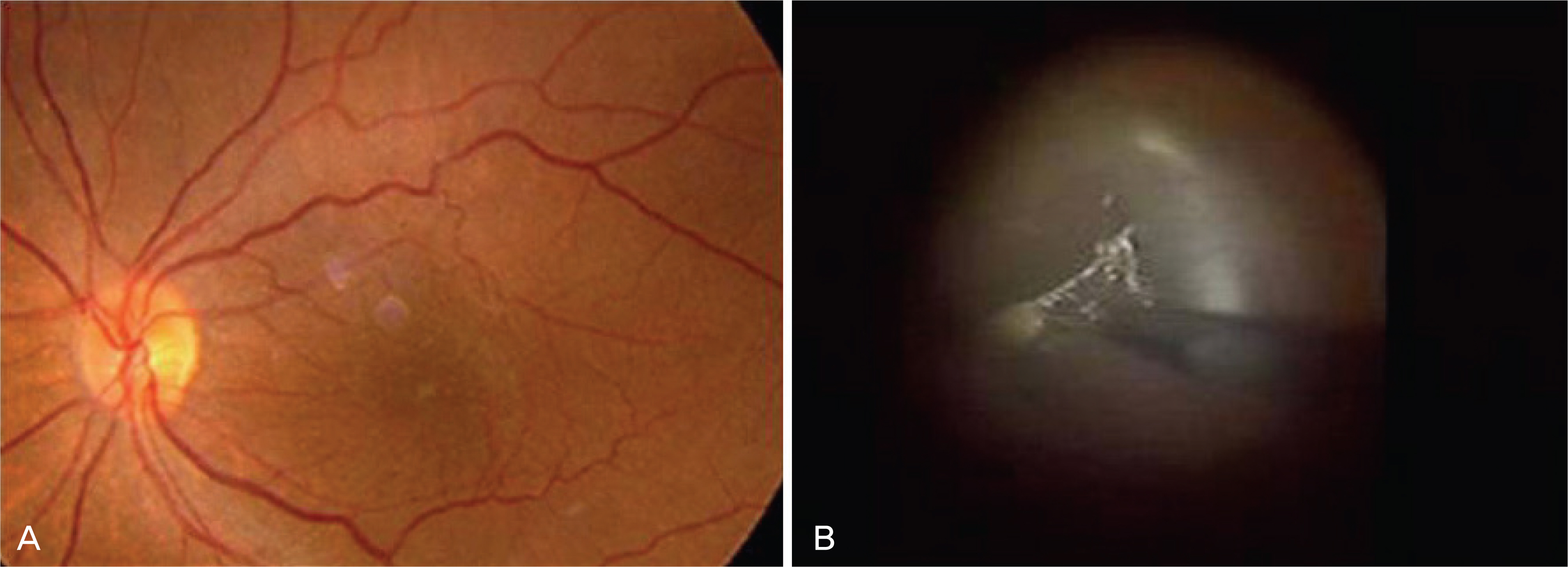
Figure 1. (A) Preoperative color photography showed thin macular epiretinal membrane. (B) Intraoperative photograph shows internal limiting membrane is peeled off with microforceps from an area of approximately 3DD surrounding the fovea.
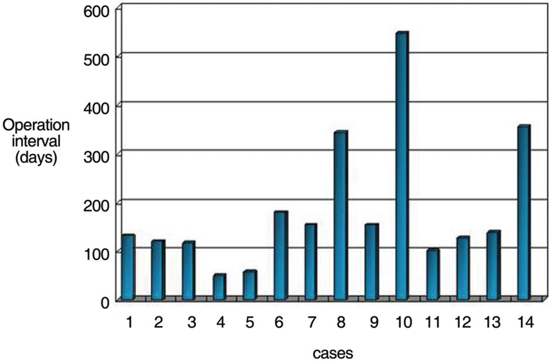
Figure 2. Time interval between retinal detachment surgery and removal of epiretinal membrane.
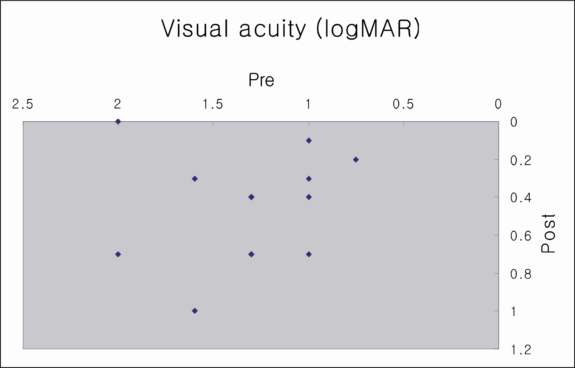
Figure 3. Preoperative and postoperative visual acuity of epiretinal membrane eyes.
Reference
-
References
1. Trese MT, Chandler DB, Machemer R. Epiretinal membrane. I. Prognostic criteria. Graefes Arch Clin Exp Ophthalmol. 1983; 221:12–5.2. Haritoglou C, Gandorfer A, Gass CA, et al. Trypan blue in macular pucker surgery: an evaluation of histology and functional outcome. Retina. 2004; 24:582–90.
Article3. Kraushar MF, Morse PH. The relationship between retina surgery and preretinal macular fibrosis. Ophthalmic Surg. 1988; 19:843–8.
Article4. Lobes LA Jr, Burton TC. The incidence of epiretinal membrane after retinal detachment surgery. Am J Ophthalmol. 1978; 85:72–7.5. Kim YI, Yoon IH. The electronmicroscopic feature of idiopathic and complicated epiretinal membrane. J Korean Ophthalmol Soc. 1992; 33:230–7.6. Cox MS, Azen SP, Barr CC, et al. Epiretinal membrane after successful surgery for proliferative vitreoretinopathy. Ophthalmology. 1995; 102:1884–91.7. Wise GN. Clinical features of idiopathic preretinal macular fibrosis. Schoenberg Lecture. Am J Ophthalmol. 1975; 79:349–7.8. Albert DM, Jakobiec FA. Principles and Practice of Ophthalmology. 2nd ed.3:Saunders;2000. p. 2103–10.9. Sheard RM, Sethi C, Greagor Z. Acute epiretinal membrane. Ophthalmology. 2003; 110:1178–84.10. Federman JL, Gouras P. Retina and vitreous. 1st ed.St. Louis: Mosby;1994. p. 10–11.11. Michels RG, Gilbert HS. Surgical management of macular pucker after retinal reattachment surgery. Am J Ophthalmol. 1979; 88:925–9.
Article12. Uemura A, Ideta H, Nagasaki H, et al. Epiretinal membrane after retinal detachment surgery. Ophthalmic Surg. 1992; 23:116–9.13. Bonnet M, Guenoun S. Surgical risk factors for severe postoperative proliferative vitreoretinopathy (PVR) in retinal detachment with grade B PVR. Graefes Arch Clin Exp Ophthalmol. 1995; 233:789–91.
Article14. Lewis H, Burke JM, Abrams GW, Aaberg TM. Perisilicone proliferation after vitrectomy for proliferative vitreoretinopathy. Ophthalmology. 1988; 95:583–91.
Article15. Shea M. The surgical management of epiretinal membrane in rhegmatogenous retinal detachment. Ophthalmology. 1980; 87:70–4.16. Hagler WS, Aturaliya U. Epiretinal membrane after retinal detachment surgery. Br J Ophthalmol. 1971; 55:451–7.17. De Bustros S, Rice TA, Michels RD, et al. Vitrectomy for epiretinal membrane. Use after treatment for retinal tears or retinal detachment. Arch Ophthalmol. 1988; 106:758–60.18. Rice TA, De Bustros S, Michels RG, et al. Prognostic factors in vitrectomy for epiretinal membranes of the macula. Ophthalmology. 1986; 93:602–10.
Article19. Poliner LS, Olk RJ, Grand MG, et al. The surgical management of premacular fibroplasia. Arch Ophthalmol. 1988; 106:761–4.20. Park SJ, Lee JH. Clinical feature of macular preretinal membrane and visual changes after vitrectomy. J Korean Ophthalmol Soc. 1994; 35:824–9.21. Lee HJ, Kim HC. The clinical outcome and prognostic factors of vitrectomy for macular epiretinal membranes. J Korean Ophthalmol Soc. 2003; 44:655–62.
- Full Text Links
-
- Actions
-
Cited
- CITED
-
- Close
- Share
-
- Similar articles
-
- The Electron Microscopic Feature of Idiopathic and Complicated Epiretinal Membrane
- Electron Microscopic Features of Epiretinal Membrane in Rhegmatogenous Retinal Detachment
- Analysis of Leading Diseases Causing Epiretinal Membrane and Comparison of Prognosis after Epiretinal Membrane Peeling
- Spontaneous Disappearance of A Traumatic Macular Hole
- Surgical Outcome of Epiretinal Membrane and Internal Limiting Membrane Removal for Macular Hole Retinal Detachment
