Large tubular colonic duplication in an adult treated with a small midline incision
- Affiliations
-
- 1Department of Surgery, Hospital Selayang, Lebuhraya Selayang-Kepong, Selangor, Malaysia.
- 2Department of Surgery, Samsung Medical Center, Sungkyunkwan University School of Medicine, Seoul, Korea. hkchun@skku.edu
- KMID: 2212222
- DOI: http://doi.org/10.4174/jkss.2012.82.3.190
Abstract
- Tubular colonic duplication presenting in adults is rare and difficult to diagnose preoperatively. Only a few cases have been reported in the literature. We report a case of a 29-year-old lady presenting with a long history of chronic constipation, abdominal mass and repeated episodes of abdominal pain. The abdominal-pelvic computed tomography scan showed segmental bowel wall thickening thought to be small bowel, and dilatation with stasis of intraluminal content. The provisional diagnosis was small bowel duplication. She was scheduled for single port laparoscopic resection. However, a T-shaped tubular colonic duplication at sigmoid colon was found intraoperatively. Resection of the large T-shaped tubular colonic duplication containing multiple impacted large fecaloma and primary anastomosis was performed. There was no perioperative complication. We report, herein, the case of a T-shaped tubular colonic duplication at sigmoid colon in an adult who was successfully treated through mini-laparotomy assisted by single port laparoscopic surgery.
MeSH Terms
Figure
-
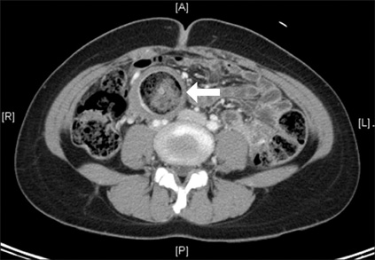
Fig. 1 Axial view abdominal computed tomography scan showing thickened bowel wall (white arrow) and segmental dilatation with stasis of intraluminal content.
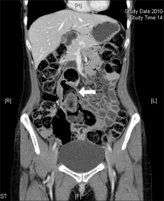
Fig. 2 Coronal view abdominal computed tomography scan showing longitudinal section of colonic duplication (white arrow).
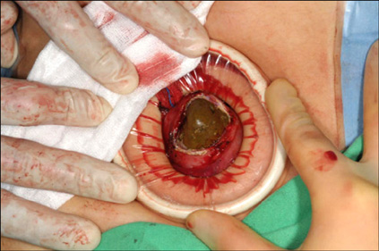
Fig. 3 Fecaloma removed through incision on colonic duplication.

Fig. 4 Sigmoid and colonic duplication extracted through wound retractor. Note that duplication arises from mesenteric border of native colon and closely wrapped around by mesocolon.
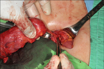
Fig. 5 Final attachment of blind end of colonic duplication to peritoneum overlying aortic bifurcation.
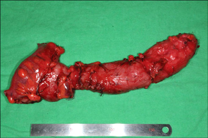
Fig. 6 Resected specimen showing large T-shaped tubular colonic duplication measuring 23 cm in length. Note that feeding vessels were ligated flush to wall of colonic duplication to avoid injury of vessels to native colon.
Reference
-
1. Macpherson RI. Gastrointestinal tract duplications: clinical, pathologic, etiologic, and radiologic considerations. Radiographics. 1993. 13:1063–1080.2. Fotiadis C, Genetzakis M, Papandreou I, Misiakos EP, Agapitos E, Zografos GC. Colonic duplication in adults: report of two cases presenting with rectal bleeding. World J Gastroenterol. 2005. 11:5072–5074.3. Yousefzadeh DK, Bickers GH, Jackson JH Jr, Benton C. Tubular colonic duplication - review of 1876-1981 literature. Pediatr Radiol. 1983. 13:65–71.4. Park YA, Jung EJ, Han SJ. Laparoscopic resection of duplicated sigmoid colon under the guidance of intraoperative colonoscopy. Surg Laparosc Endosc Percutan Tech. 2005. 15:299–301.5. Payne CE, Deshon GE Jr, Kroll JD, Sumfest J. Colonic duplication: an unusual cause of enterovesical fistula. Urology. 1995. 46:726–728.6. Odofin O, Yianni L, Al-Talibi A, Gore DM. Perforation of an ileal duplication presenting as an acute abdomen. Surgeon. 2010. 8:117–118.7. Lee J, Jeon YH, Lee S. Papillary adenocarcinoma arising in a duplication of the cecum. Abdom Imaging. 2008. 33:601–603.8. Kume K, Sakata H, Otsuki M. Education and imaging. Gastrointestinal: tubular duplication of the descending colon. J Gastroenterol Hepatol. 2007. 22:1553.9. Carneiro FP, de Nazareth Sobreira M, de Azevedo AE, Alves AP, Campos KM. Colonic duplication in an adult mimicking a tumor of pancreas. World J Gastroenterol. 2008. 14:966–968.10. Lim GY, Im SA, Chung JH. Complicated duplication cysts on the ileum presenting with a mesenteric inflammatory mass. Pediatr Radiol. 2008. 38:467–470.
