Clinical treatment of postoperative infection following sinus augmentation
- Affiliations
-
- 1Gangnam Hyundai Dental Clinic, Seoul, Korea.
- 2Luden Dental Clinic, Seoul, Korea.
- 3Department of Periodontology, Kyung Hee University School of Dentistry, Seoul, Korea.
- 4Department of Dentistry/Periodontology, Hanyang University School of Medicine, Seoul, Korea.
- 5Department of Periodontology, Institute of Oral Biology, Kyung Hee University School of Dentistry, Seoul, Korea. chungjh@khu.ac.kr
- KMID: 2212142
- DOI: http://doi.org/10.5051/jpis.2010.40.3.144
Abstract
- PURPOSE
The aim of this case report is to present the successful clinical treatment of two cases of postoperative infection following maxillary sinus augmentation.
METHODS
In the two cases of postoperative infection, immediate total removal of the grafted material from the sinus was conducted to stop the spread of the infection, after which a high dose of antibiotics was administrated. Re-augmentation procedures were then conducted after the infection subsided.
RESULTS
No further complications occurred after sinus re-augmentation. The dental implants placed in the re-augmented sinus were clinically osseointegrated, and the implant-supported restorations in the two cases of postoperative infection have been functioning very well for over 2 years.
CONCLUSIONS
In the case of infection of the grafted sinuses, it is necessary to completely remove the graft materials and then administer a high dose of antibiotics to treat the acute infection, after which sinus re-augmentation is suggested.
MeSH Terms
Figure
-
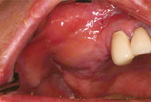
Figure 1 Postoperative infection following sinus augmentation.
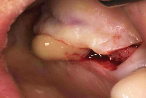
Figure 2 The discharge of pus and graft materials after the incision.

Figure 3 Postoperative panoramic radiograph following sinus re-augmentation along with the placement of dental implants.
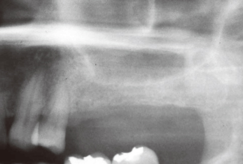
Figure 4 Pneumatization on the left maxillary sinus was observed in the preoperative panoramic radiograph.
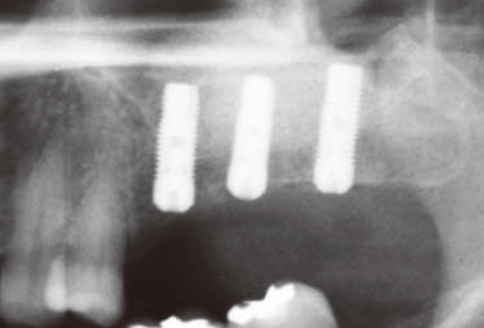
Figure 5 Implant placed on positions #25-27 simultaneously with the sinus augmentation.
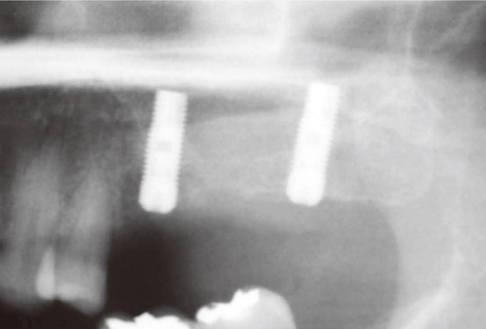
Figure 6 Infected graft material was surgically removed and #26i was removed to ease removal of the infected graft.

Figure 7 The inner portion of the buccal flap and the sinus membrane were fused. The membrane and the residual graft material were inseparably fused together.
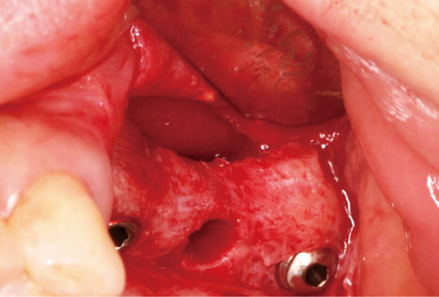
Figure 8 Elevation of the sinus membrane and ostectomy were performed.
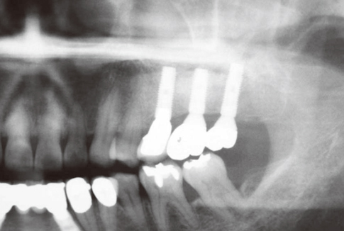
Figure 9 Panoramic radiograph of the dental implants placed in the re-grafted sinus after successfully functioning for 33 months.
Cited by 1 articles
-
Location of maxillary intraosseous vascular anastomosis based on the tooth position and height of the residual alveolar bone: computed tomographic analysis
Seung-Min Yang, Seung-Beom Kye
J Periodontal Implant Sci. 2014;44(2):50-56. doi: 10.5051/jpis.2014.44.2.50.
Reference
-
1. Albrektsson T. A multicenter report on osseointegrated oral implants. J Prosthet Dent. 1988. 60:75–84.
Article2. Engquist B, Bergendal T, Kallus T, Linden U. A retrospective multicenter evaluation of osseointegrated implants supporting overdentures. Int J Oral Maxillofac Implants. 1988. 3:129–134.3. Jemt T, Lekholm U, Adell R. Osseointegrated implants in the treatment of partially edentulous patients: a preliminary study on 876 consecutively placed fixtures. Int J Oral Maxillofac Implants. 1989. 4:211–217.4. Boyne PJ, James RA. Grafting of the maxillary sinus floor with autogenous marrow and bone. J Oral Surg. 1980. 38:613–616.5. Misch CE. Maxillary sinus augmentation for endosteal implants: organized alternative treatment plans. Int J Oral Implantol. 1987. 4:49–58.6. Summers RB. The osteotome technique: Part 3--Less invasive methods of elevating the sinus floor. Compendium. 1994. 15:698–710.7. Fugazzotto PA, De PS. Sinus floor augmentation at the time of maxillary molar extraction: success and failure rates of 137 implants in function for up to 3 years. J Periodontol. 2002. 73:39–44.
Article8. Wallace SS, Froum SJ. Effect of maxillary sinus augmentation on the survival of endosseous dental implants. A systematic review. Ann Periodontol. 2003. 8:328–343.
Article9. Mardinger O, Nissan J, Chaushu G. Sinus floor augmentation with simultaneous implant placement in the severely atrophic maxilla: technical problems and complications. J Periodontol. 2007. 78:1872–1877.
Article10. Hernandez-Alfaro F, Torradeflot MM, Marti C. Prevalence and management of Schneiderian membrane perforations during sinus-lift procedures. Clin Oral Implants Res. 2008. 19:91–98.
Article11. Pikos MA. Maxillary sinus membrane repair: update on technique for large and complete perforations. Implant Dent. 2008. 17:24–31.
Article12. Ardekian L, Oved-Peleg E, Mactei EE, Peled M. The clinical significance of sinus membrane perforation during augmentation of the maxillary sinus. J Oral Maxillofac Surg. 2006. 64:277–282.
Article13. Lockhart R, Ceccaldi J, Bertrand JC. Postoperative maxillary cyst following sinus bone graft: report of a case. Int J Oral Maxillofac Implants. 2000. 15:583–586.14. Misch CE. Contemporary implant dentistry. 2008. 3rd ed. St. Louis: Mosby/Elsevier.15. Barone A, Santini S, Sbordone L, Crespi R, Covani U. A clinical study of the outcomes and complications associated with maxillary sinus augmentation. Int J Oral Maxillofac Implants. 2006. 21:81–85.16. Schwartz-Arad D, Herzberg R, Dolev E. The prevalence of surgical complications of the sinus graft procedure and their impact on implant survival. J Periodontol. 2004. 75:511–516.
Article17. Zijderveld SA, van den Bergh JP, Schulten EA, ten Bruggenkate CM. Anatomical and surgical findings and complications in 100 consecutive maxillary sinus floor elevation procedures. J Oral Maxillofac Surg. 2008. 66:1426–1438.
Article18. Anavi Y, Allon DM, Avishai G, Calderon S. Complications of maxillary sinus augmentations in a selective series of patients. Oral Surg Oral Med Oral Pathol Oral Radiol Endod. 2008. 106:34–38.
Article19. Misch CM. The pharmacologic management of maxillary sinus elevation surgery. J Oral Implantol. 1992. 18:15–23.20. Lindhe J, Lang NP, Karring T. Clinical periodontology and implant dentistry. 2008. Oxford: Blackwell Munksgaard.21. Bravetti P, Membre H, Marchal L, Jankowski R. Histologic changes in the sinus membrane after maxillary sinus augmentation in goats. J Oral Maxillofac Surg. 1998. 56:1170–1176.
Article22. Sul SH, Choi BH, Li J, Jeong SM, Xuan F. Histologic changes in the maxillary sinus membrane after sinus membrane elevation and the simultaneous insertion of dental implants without the use of grafting materials. Oral Surg Oral Med Oral Pathol Oral Radiol Endod. 2008. 105:e1–e5.
Article
- Full Text Links
-
- Actions
-
Cited
- CITED
-
- Close
- Share
-
- Similar articles
-
- Management of Perioperative Pathologic Conditions Involving Maxillary Sinus for Dental Implant Placement and Sinus Augmentation: Report of Case Series and Literature Review
- Sinus floor augmentation at the time of tooth removal
- Delayed Occurrence of Maxillary Sinusitis after Simultaneous Maxillary Sinus Augmentation and Implant: A Case Report and Literature Review
- A Case of Maxillary Sinusitis after Sinus Floor Augmentation
- A review of complications of maxillary sinus augmentation and available treatment methods
