Bone regeneration effects of human allogenous bone substitutes: a preliminary study
- Affiliations
-
- 1Department of Periodontology and Dental Research Institute, Seoul National University College of Dentistry, Seoul, Korea. periopf@snu.ac.kr
- KMID: 2212140
- DOI: http://doi.org/10.5051/jpis.2010.40.3.132
Abstract
- PURPOSE
The purpose of this study was to compare the bone regeneration effects of cortical, cancellous, and cortico-cancellous human bone substitutes on calvarial defects of rabbits.
METHODS
Four 8-mm diameter calvarial defects were created in each of nine New Zealand white rabbits. Freeze-dried cortical bone, freeze-dried cortico-cancellous bone, and demineralized bone matrix with freeze-dried cancellous bone were inserted into the defects, while the non-grafted defect was regarded as the control. After 4, 8, and 12 weeks of healing, the experimental animals were euthanized for specimen preparation. Micro-computed tomography (micro-CT) was performed to calculate the percent bone volume. After histological evaluation, histomorphometric analysis was performed to quantify new bone formation.
RESULTS
In micro-CT evaluation, freeze-dried cortico-cancellous human bone showed the highest percent bone volume value among the experimental groups at week 4. At week 8 and week 12, freeze-dried cortical human bone showed the highest percent bone volume value among the experimental groups. In histologic evaluation, at week 4, freeze-dried cortico-cancellous human bone showed more prominent osteoid tissue than any other group. New bone formation was increased in all of the experimental groups at week 8 and 12. Histomorphometric data showed that freeze-dried cortico-cancellous human bone showed a significantly higher new bone formation percentile value than any other experimental group at week 4. At week 8, freeze-dried cortical human bone showed the highest value, of which a significant difference existed between freeze-dried cortical human bone and demineralized bone matrix with freeze-dried cancellous human bone. At week 12, there were no significant differences among the experimental groups.
CONCLUSIONS
Freeze-dried cortico-cancellous human bone showed swift new bone formation at the 4-week healing phase, whereas there was less difference in new bone formation among the experimental groups in the following healing phases.
MeSH Terms
Figure
-
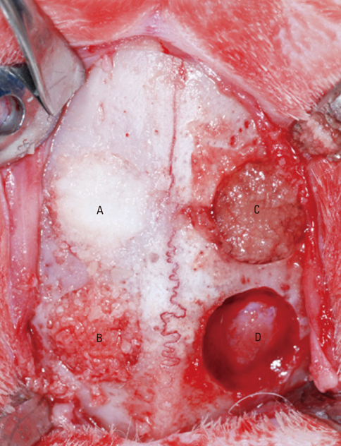
Figure 1 Four 8-mm diameter calvarial defects were created. Freeze-dried cortical human bone (A), freeze-dried cortico-cancellous human bone (B), and demineralized bone matrix with cancellous human bone (C) were inserted into the three experimental defect sites. No biomaterial was grafted into the control defect site (D).
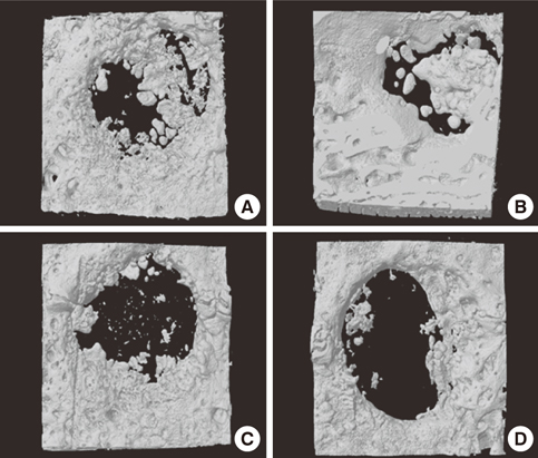
Figure 2 Three dimensional images of freeze-dried cortical human bone (A), freeze-dried cortico-cancellous human bone (B), demineralized bone matrix with cancellous human bone (C), and the control (D) at week 4. Freeze-dried cortico-cancellous human bone (B) revealed greatest amount of newly formed bone.
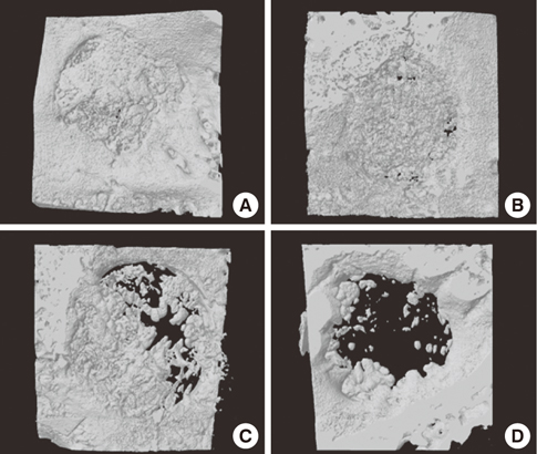
Figure 3 Three dimensional images of freeze-dried cortical human bone (A), freeze-dried cortico-cancellous human bone (B), demineralized bone matrix with cancellous human bone (C), and the control (D) at week 8. Freeze-dried cortical human bone (A) showed better development of trabecular bone than any other group.
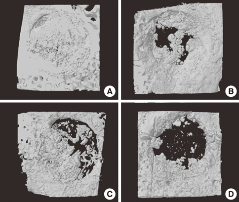
Figure 4 Three dimensional images of freeze-dried cortical human bone (A), freeze-dried cortico-cancellous human bone (B), demineralized bone matrix with cancellous human bone (C), and the control (D) at week 12. The defects were nearly closed by newly formed bone, except for the control.
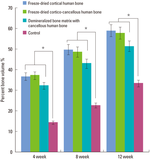
Figure 5 Percent bone volume values of each group from micro-CT analysis. All experimental groups showed significantly higher percent bone volume values than the control group at week 4, 8, and 12. *Significant difference between the experimental and the control group (P < 0.05).
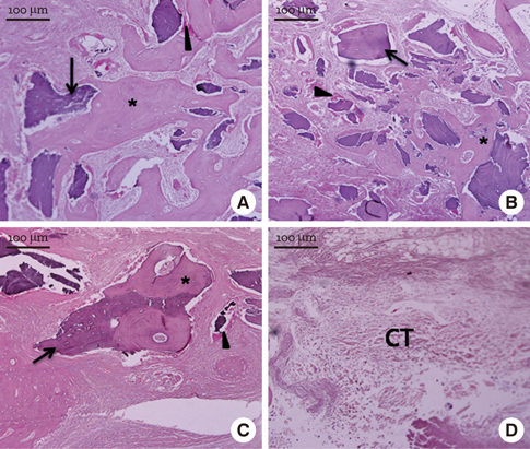
Figure 6 Light micrographs at week 4 (H&E stain). Freeze-dried cortical human bone (A), freeze-dried cortico-cancellous human bone (B), demineralized bone matrix with cancellous human bone (C), and the control (D). All experimental groups showed new bone formation (asterisk) at the margins of the defect sites. Graft materials (arrow) were surrounded by newly formed osteoid tissue (arrow-head). In freeze-dried cortico-cancellous human bone (B), newly formed bone was observed more prominently than in any other group. In the control, loose connective tissue (CT) filled the defect sites.
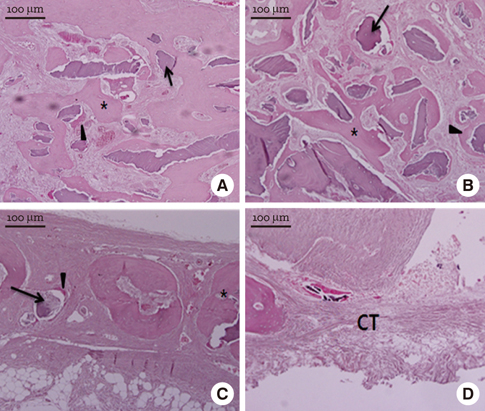
Figure 7 Light micrographs at week 8 (H&E stain). Freeze-dried cortical human bone (A), freeze-dried cortico-cancellous human bone (B), demineralized bone matrix with cancellous human bone (C), and the control (D). The new bone (asterisk) surrounded the graft particles (arrow). There were abundant osteogenic cells and matrix (arrowhead) around the graft particles. In the control, the central area was still filled mainly with loose connective tissue (CT).
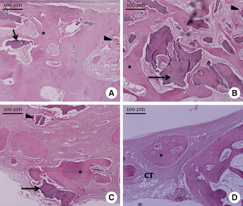
Figure 8 Light micrographs at week 12 (H&E stain). Freeze-dried cortical human bone (A), freeze-dried cortico-cancellous human bone (B), demineralized bone matrix with cancellous human bone (C), and the control (D). The newly formed bone (asterisk) showed a more mature pattern than at week 4 or 8. In the control, loose connective tissue (CT) still filled most of the defect area.
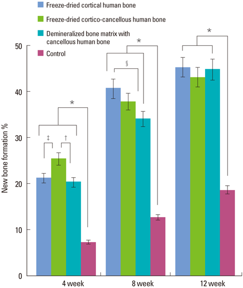
Figure 9 New bone formation values of each group from histomorphometric analysis. All experimental groups showed significantly higher new bone formation values than the control at week 4, 8, and 12. *Significant difference between the experimental and the control groups (P < 0.05). †Significant difference between freeze-dried cortico-cancellous human bone and demineralized bone matrix with cancellous human bone (P < 0.05). ‡Significant difference between freeze-dried cortical human bone and freeze-dried cortico-cancellous human bone (P < 0.05). §Significant difference between freeze-dried cortical human bone and demineralized bone matrix having cancellous human bone (P < 0.05).
Reference
-
1. Helm GA, Dayoub H, Jane JA Jr. Bone graft substitutes for the promotion of spinal arthrodesis. Neurosurg Focus. 2001; 10:E4.
Article2. Giannoudis PV, Dinopoulos H, Tsiridis E. Bone substitutes: an update. Injury. 2005; 36:Suppl 3. S20–S27.
Article3. Urist MR. Bone: formation by autoinduction. Science. 1965; 150:893–899.
Article4. Misch CE, Dietsh F. Bone-grafting materials in implant dentistry. Implant Dent. 1993; 2:158–167.
Article5. Pietrzak WS, Woodell-May J, McDonald N. Assay of bone morphogenetic protein-2, -4, and -7 in human demineralized bone matrix. J Craniofac Surg. 2006; 17:84–90.
Article6. Mellonig JT, Bowers GM, Bailey RC. Comparison of bone graft materials. Part I. New bone formation with autografts and allografts determined by Strontium-85. J Periodontol. 1981; 52:291–296.
Article7. Mellonig JT, Bowers GM, Cotton WR. Comparison of bone graft materials. Part II. New bone formation with autografts and allografts: a histological evaluation. J Periodontol. 1981; 52:297–302.
Article8. Piattelli A, Scarano A, Corigliano M, Piattelli M. Comparison of bone regeneration with the use of mineralized and demineralized freeze-dried bone allografts: a histological and histochemical study in man. Biomaterials. 1996; 17:1127–1131.
Article9. Rummelhart JM, Mellonig JT, Gray JL, Towle HJ. A comparison of freeze-dried bone allograft and demineralized freeze-dried bone allograft in human periodontal osseous defects. J Periodontol. 1989; 60:655–663.
Article10. Mellonig JT, Bowers GM, Bright RW, Lawrence JJ. Clinical evaluation of freeze-dried bone allografts in periodontal osseous defects. J Periodontol. 1976; 47:125–131.
Article11. Cammack GV 2nd, Nevins M, Clem DS 3rd, Hatch JP, Mellonig JT. Histologic evaluation of mineralized and demineralized freeze-dried bone allograft for ridge and sinus augmentations. Int J Periodontics Restorative Dent. 2005; 25:231–237.12. Cornell CN. Osteobiologics. Bull Hosp Jt Dis. 2004; 62:13–17.13. Behairy Y, Jasty M. Bone grafts and bone substitutes in hip and knee surgery. Orthop Clin North Am. 1999; 30:661–671.
Article14. Goldberg VM, Stevenson S. Natural history of autografts and allografts. Clin Orthop Relat Res. 1987; 7–16.
Article15. Ludwig SC, Boden SD. Osteoinductive bone graft substitutes for spinal fusion: a basic science summary. Orthop Clin North Am. 1999; 30:635–645.16. Schreurs BW, Slooff TJ, Buma P, Gardeniers JW, Huiskes R. Acetabular reconstruction with impacted morsellised cancellous bone graft and cement. A 10- to 15-year follow-up of 60 revision arthroplasties. J Bone Joint Surg Br. 1998; 80:391–395.
- Full Text Links
-
- Actions
-
Cited
- CITED
-
- Close
- Share
-
- Similar articles
-
- The comparative study of bone substitute materials in bone regeneration
- Biomaterial development for oral and maxillofacial bone regeneration
- Hard tissue regeneration using bone substitutes: an update on innovations in materials
- Bone Substitutes and the Advancement for Enhancing Bone Healing
- Effect of deproteinized bovine bone mineral on cell proliferation in the procedure of guided bone regeneration
