Accuracy of computer-aided template-guided oral implant placement: a prospective clinical study
- Affiliations
-
- 1Department of Dental Implants, U.O.C. Maxillofacial Surgery and Odontostomatology, Fondazione IRCCS Ca Granda, State University of Milan, Milan, Italy. pierpaolo.poli@unimi.it
- KMID: 2212012
- DOI: http://doi.org/10.5051/jpis.2014.44.4.184
Abstract
- PURPOSE
The aim of the present study was to evaluate the in vivo accuracy of flapless, computer-aided implant placement by comparing the three-dimensional (3D) position of planned and placed implants through an analysis of linear and angular deviations.
METHODS
Implant position was virtually planned using 3D planning software based on the functional and aesthetic requirements of the final restorations. Computer-aided design/computer-assisted manufacture technology was used to transfer the virtual plan to the surgical environment. The 3D position of the planned and placed implants, in terms of the linear deviations of the implant head and apex and the angular deviations of the implant axis, was compared by overlapping the pre- and postoperative computed tomography scans using dedicated software.
RESULTS
The comparison of 14 implants showed a mean linear deviation of the implant head of 0.56 mm (standard deviation [SD], 0.23), a mean linear deviation of the implant apex of 0.64 mm (SD, 0.29), and a mean angular deviation of the long axis of 2.42degrees (SD, 1.02).
CONCLUSIONS
In the present study, computer-aided flapless implant surgery seemed to provide several advantages to the clinicians as compared to the standard procedure; however, linear and angular deviations are to be expected. Therefore, accurate presurgical planning taking into account anatomical limitations and prosthetic demands is mandatory to ensure a predictable treatment, without incurring possible intra- and postoperative complications.
MeSH Terms
Figure
-

Figure 1 Preoperative frontal view of the edentulous jaws.

Figure 2 Computer-aided planning in the upper jaw.
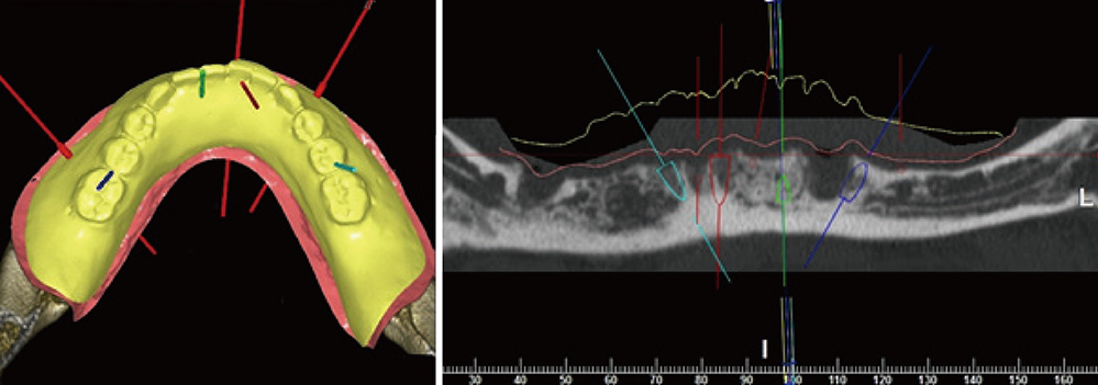
Figure 3 Computer-aided planning in the lower jaw.
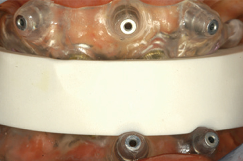
Figure 4 Stabilization of the two surgical stent by means of a silicone index.
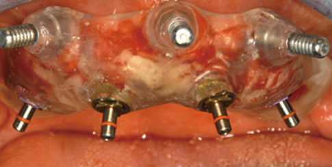
Figure 5 Upper surgical stent fixed in the proper position by means of cortical pins and implants insertion.
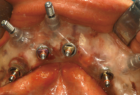
Figure 6 Lower surgical stent fixed in the proper position by means of cortical pins and implants insertion.
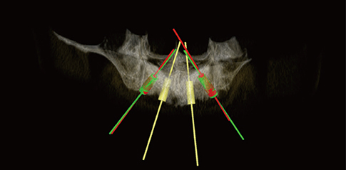
Figure 7 Overlapping of the pre- and postoperative scans for the comparison between planned (red) and real (green) implant positions in order to analyse the accuracy of the system in the upper jaw.
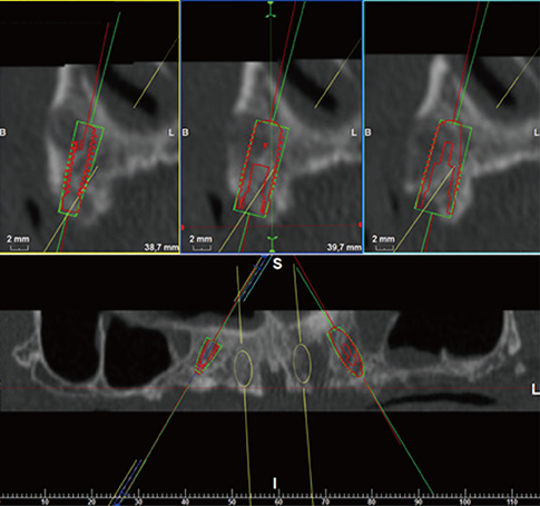
Figure 8 Accuracy assessments in the upper jaw between planned (red) and real (green) implant positions.

Figure 9 Overlapping of the pre and postoperative scans for the comparison between planned (red) and real (green) implant positions in order to analyse the accuracy of the system in the lower jaw.
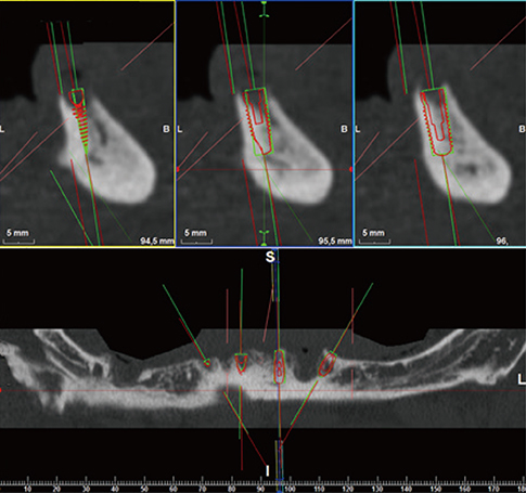
Figure 10 Accuracy assessments in the lower jaw between planned (red) and real (green) implant positions.
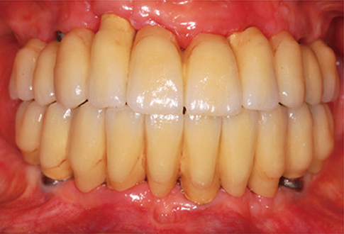
Figure 11 Frontal view of the final upper and lower restorations.
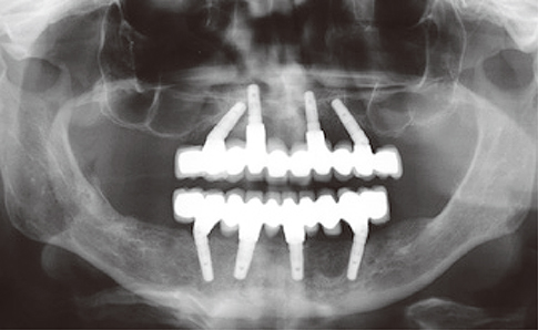
Figure 12 Follow-up X-rays control.
Reference
-
1. Rangert B, Krogh PH, Langer B, Van Roekel N. Bending overload and implant fracture: a retrospective clinical analysis. Int J Oral Maxillofac Implants. 1995; 10:326–334.2. Hobkirk JA, Havthoulas TK. The influence of mandibular deformation, implant numbers, and loading position on detected forces in abutments supporting fixed implant superstructures. J Prosthet Dent. 1998; 80:169–174.
Article3. Stanford CM. Biomechanical and functional behavior of implants. Adv Dent Res. 1999; 13:88–92.
Article4. Schwartz-Arad D, Levin L. Intraoral autogenous block onlay bone grafting for extensive reconstruction of atrophic maxillary alveolar ridges. J Periodontol. 2005; 76:636–641.
Article5. Maiorana C, Santoro F. Maxillary and mandibular bone reconstruction with hip grafts and implants using Frialit-2 implants. Int J Periodontics Restorative Dent. 2002; 22:221–229.6. Simion M, Trisi P, Piattelli A. Vertical ridge augmentation using a membrane technique associated with osseointegrated implants. Int J Periodontics Restorative Dent. 1994; 14:496–511.7. Chiapasco M, Lang NP, Bosshardt DD. Quality and quantity of bone following alveolar distraction osteogenesis in the human mandible. Clin Oral Implants Res. 2006; 17:394–402.
Article8. Engelke WG, Diederichs CG, Jacobs HG, Deckwer I. Alveolar reconstruction with splitting osteotomy and microfixation of implants. Int J Oral Maxillofac Implants. 1997; 12:310–318.9. Becker CM, Kaiser DA. Surgical guide for dental implant placement. J Prosthet Dent. 2000; 83:248–251.
Article10. Almog DM, Torrado E, Meitner SW. Fabrication of imaging and surgical guides for dental implants. J Prosthet Dent. 2001; 85:504–508.
Article11. Garber DA. The esthetic dental implant: letting restoration be the guide. J Am Dent Assoc. 1995; 126:319–325.
Article12. Pesun IJ, Gardner FM. Fabrication of a guide for radiographic evaluation and surgical placement of implants. J Prosthet Dent. 1995; 73:548–552.
Article13. Di Giacomo GA, Cury PR, de Araujo NS, Sendyk WR, Sendyk CL. Clinical application of stereolithographic surgical guides for implant placement: preliminary results. J Periodontol. 2005; 76:503–507.
Article14. Sarment DP, Al-Shammari K, Kazor CE. Stereolithographic surgical templates for placement of dental implants in complex cases. Int J Periodontics Restorative Dent. 2003; 23:287–295.15. van Steenberghe D, Naert I, Andersson M, Brajnovic I, Van Cleynenbreugel J, Suetens P. A custom template and definitive prosthesis allowing immediate implant loading in the maxilla: a clinical report. Int J Oral Maxillofac Implants. 2002; 17:663–670.16. Sarment DP, Sukovic P, Clinthorne N. Accuracy of implant placement with a stereolithographic surgical guide. Int J Oral Maxillofac Implants. 2003; 18:571–577.17. Van Assche N, van Steenberghe D, Guerrero ME, Hirsch E, Schutyser F, Quirynen M, et al. Accuracy of implant placement based on pre-surgical planning of three-dimensional cone-beam images: a pilot study. J Clin Periodontol. 2007; 34:816–821.
Article18. Fortin T, Champleboux G, Lormee J, Coudert JL. Precise dental implant placement in bone using surgical guides in conjunction with medical imaging techniques. J Oral Implantol. 2000; 26:300–303.
Article19. Fortin T, Champleboux G, Bianchi S, Buatois H, Coudert JL. Precision of transfer of preoperative planning for oral implants based on cone-beam CT-scan images through a robotic drilling machine. Clin Oral Implants Res. 2002; 13:651–656.
Article20. Klein M, Abrams M. Computer-guided surgery utilizing a computer-milled surgical template. Pract Proced Aesthet Dent. 2001; 13:165–169.21. Widmann G, Widmann R, Widmann E, Jaschke W, Bale R. Use of a surgical navigation system for CT-guided template production. Int J Oral Maxillofac Implants. 2007; 22:72–78.22. Valente F, Schiroli G, Sbrenna A. Accuracy of computer-aided oral implant surgery: a clinical and radiographic study. Int J Oral Maxillofac Implants. 2009; 24:234–242.23. Ersoy AE, Turkyilmaz I, Ozan O, McGlumphy EA. Reliability of implant placement with stereolithographic surgical guides generated from computed tomography: clinical data from 94 implants. J Periodontol. 2008; 79:1339–1345.
Article24. D'haese J, Van De Velde T, Komiyama A, Hultin M, De Bruyn H. Accuracy and complications using computer-designed stereolithographic surgical guides for oral rehabilitation by means of dental implants: a review of the literature. Clin Implant Dent Relat Res. 2012; 14:321–335.25. Jung RE, Schneider D, Ganeles J, Wismeijer D, Zwahlen M, Hammerle CH, et al. Computer technology applications in surgical implant dentistry: a systematic review. Int J Oral Maxillofac Implants. 2009; 24:92–109.
Article26. Tahmaseb A, Wismeijer D, Coucke W, Derksen W. Computer technology applications in surgical implant dentistry: a systematic review. Int J Oral Maxillofac Implants. 2014; 29:25–42.
Article27. De Bruyn H, Atashkadeh M, Cosyn J, van de Velde T. Clinical outcome and bone preservation of single TiUnite™ implants installed with flapless or flap surgery. Clin Implant Dent Relat Res. 2011; 13:175–183.
Article28. Jensen OT, Cullum DR, Baer D. Marginal bone stability using 3 different flap approaches for alveolar split expansion for dental implants: a 1-year clinical study. J Oral Maxillofac Surg. 2009; 67:1921–1930.
Article29. Wood DL, Hoag PM, Donnenfeld OW, Rosenfeld LD. Alveolar crest reduction following full and partial thickness flaps. J Periodontol. 1972; 43:141–144.
Article30. Rousseau P. Flapless and traditional dental implant surgery: an open, retrospective comparative study. J Oral Maxillofac Surg. 2010; 68:2299–2306.
Article31. Brodala N. Flapless surgery and its effect on dental implant outcomes. Int J Oral Maxillofac Implants. 2009; 24:118–125.32. Sclar AG. Guidelines for flapless surgery. J Oral Maxillofac Surg. 2007; 65:7 Suppl 1. 20–32.
Article33. Pettersson A, Kero T, Gillot L, Cannas B, Faldt J, Soderberg R, et al. Accuracy of CAD/CAM-guided surgical template implant surgery on human cadavers: Part I. J Prosthet Dent. 2010; 103:334–342.
Article34. Widmann G, Zangerl A, Keiler M, Stoffner R, Bale R, Puelacher W. Flapless implant surgery in the edentulous jaw based on three fixed intraoral reference points and image-guided surgical templates: accuracy in human cadavers. Clin Oral Implants Res. 2010; 21:835–841.
Article35. Schneider D, Marquardt P, Zwahlen M, Jung RE. A systematic review on the accuracy and the clinical outcome of computer-guided template-based implant dentistry. Clin Oral Implants Res. 2009; 20:Suppl 4. 73–86.
Article36. Van Assche N, van Steenberghe D, Quirynen M, Jacobs R. Accuracy assessment of computer-assisted flapless implant placement in partial edentulism. J Clin Periodontol. 2010; 37:398–403.
Article37. Ozan O, Turkyilmaz I, Ersoy AE, McGlumphy EA, Rosenstiel SF. Clinical accuracy of 3 different types of computed tomography-derived stereolithographic surgical guides in implant placement. J Oral Maxillofac Surg. 2009; 67:394–401.
Article38. Widmann G, Bale RJ. Accuracy in computer-aided implant surgery: a review. Int J Oral Maxillofac Implants. 2006; 21:305–313.39. Cassetta M, Stefanelli LV, Giansanti M, Di Mambro A, Calasso S. Depth deviation and occurrence of early surgical complications or unexpected events using a single stereolithographic surgi-guide. Int J Oral Maxillofac Surg. 2011; 40:1377–1387.
Article40. D'haese J, Van De Velde T, Elaut L, De Bruyn H. A prospective study on the accuracy of mucosally supported stereolithographic surgical guides in fully edentulous maxillae. Clin Implant Dent Relat Res. 2012; 14:293–303.
- Full Text Links
-
- Actions
-
Cited
- CITED
-
- Close
- Share
-
- Similar articles
-
- Full mouth rehabilitation utilizing computer guided implant surgery and CAD/CAM
- A survey of the satisfaction of patients who have undergone implant surgery with and without employing a computer-guided implant surgical template
- Clinical Precautions for Implant Placement using Computer-guided Implant Surgical Guide: A Systematic Review
- An assessment of template-guided implant surgery in terms of accuracy and related factors
- Accuracy of the CT guided implant template by using an intraoral scanner according to the edentulous distance
