Bilateral Central Retinal Vein Occlusion in Primary Antiphospholipid Syndrome
- Affiliations
-
- 1Department of Ophthalmology, College of medicine,Chungnam National University, Daejeon, Korea. kimjy@cnu.ac.kr
- 2Chungnam National University Research Institute for Medical Sciences, Daejeon, Korea.
- KMID: 2211001
- DOI: http://doi.org/10.3341/jkos.2007.48.10.1433
Abstract
-
PURPOSE: To Report a case of bilateral central retinal vein occlusion (CRVO) caused by primary antiphospholipid syndrome.
METHODS
A 38-year-old male is referred to the department of ophthalmology for the bilateral visual loss.
RESULTS
On initial visit, both visual acuity was 0.3. Upon consider changing to fundoscopic examination, the patient was diagnosed with bilateral CRVO. We performed hematologic tests including thrombophilia examination. There were no abnormal findings on routine hematologic tests. Antinuclear antibody and rheumatoid factor were negative but anticardiolipin antibodies presented high titer, on two occasions six weeks apart. We prescribed oral aspirin and performed intravitreal bevacizumab injection under the diagnosis of bilateral CRVO in primary antiphospholipid syndrome.
CONCLUSIONS
It may be necessary to check antiphospolipid antibody in cases of bilateral CRVO in young patients without medical problem.
MeSH Terms
Figure
-
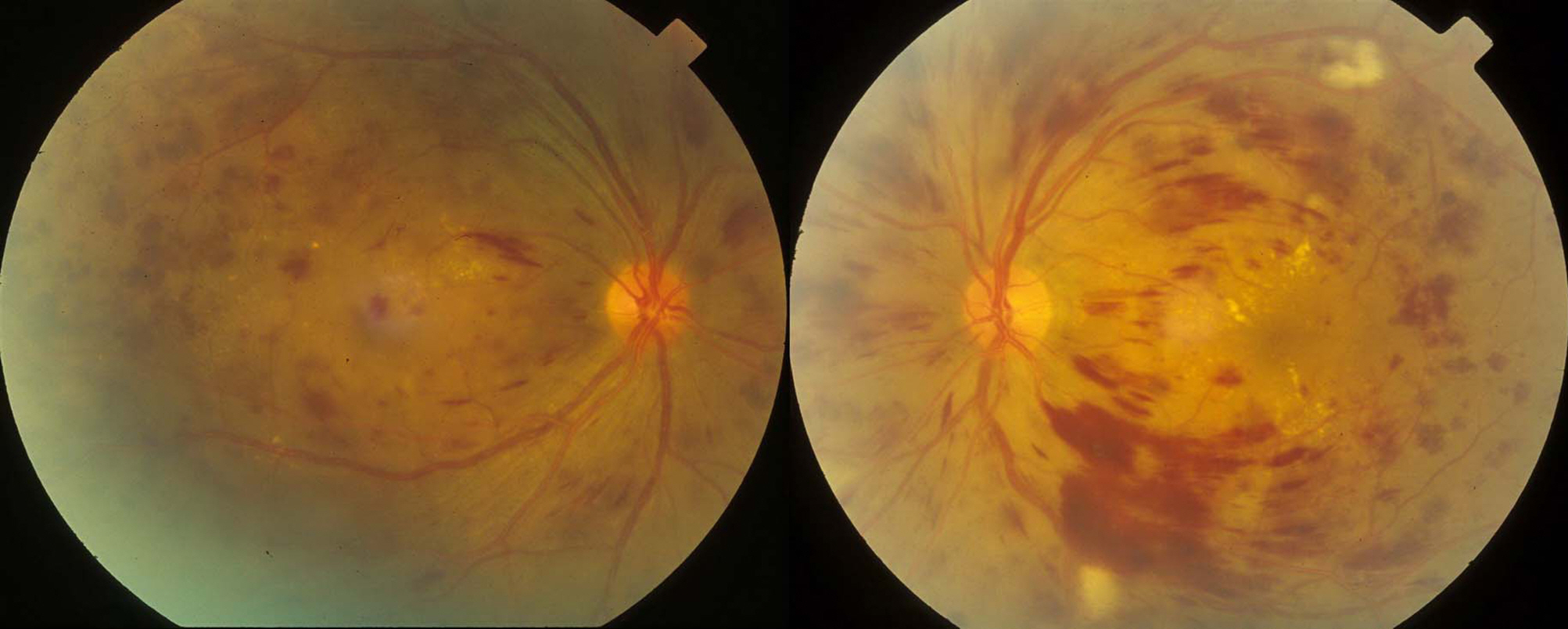
Figure 1. Fundus photographs show multiple retinal hemorrhages, macular edema, and dilated tortuous vessels.
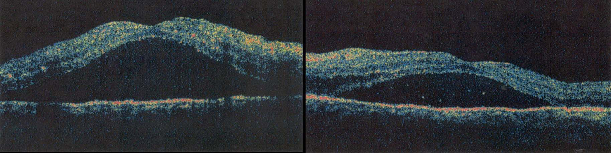
Figure 2. OCT scans show serous macular detachment in both eyes.
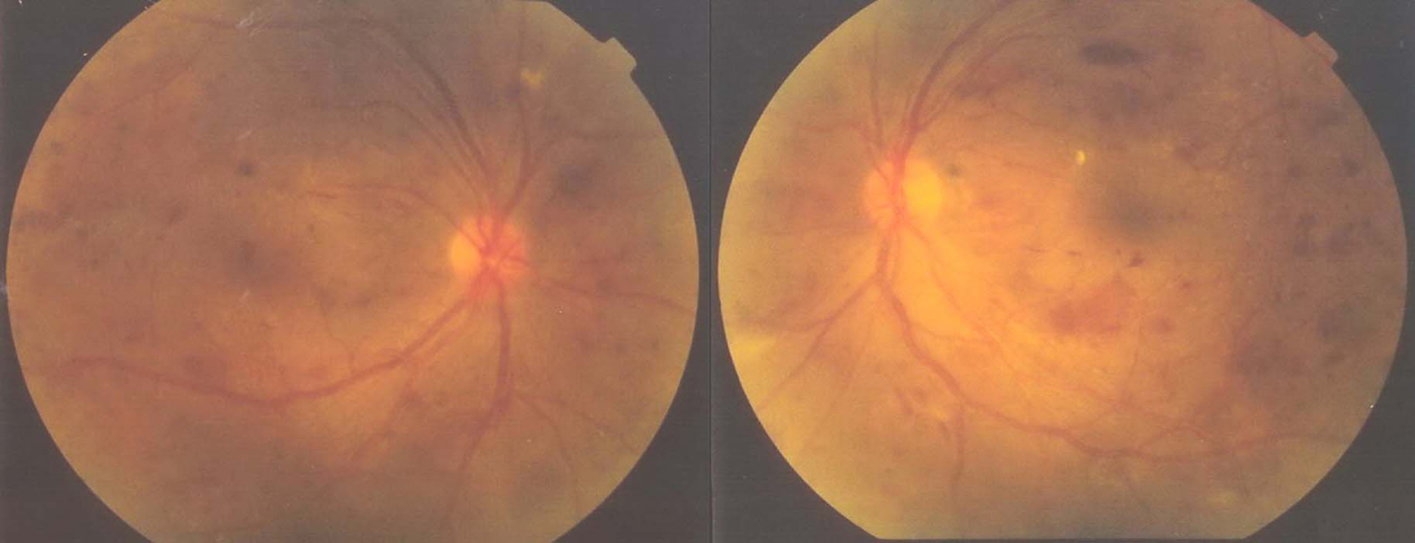
Figure 3. Fundus photographs show diminution of retinal hemorrhage and macular edema at 9 months after intravitreal bevacizumab injection.

Figure 4. OCT scans show nearly normal foveal contour and thickness at 9 months after intravitreal bevacizumab injection in both eyes.
Cited by 2 articles
-
Central Retinal Vein Occlusion in Iron Deficiency Anemia
Young Joon Jo, Deok Goo Lee, Ki Yup Nam, Jung Yeul Kim
J Korean Ophthalmol Soc. 2009;50(9):1432-1436. doi: 10.3341/jkos.2009.50.9.1432.Bilateral Central Retinal Vein Occlusion in Patient with Disseminated Intravascular Coagulation
Kyoung In Jung, Hae Ri Yum, In Tae Kim
J Korean Ophthalmol Soc. 2011;52(8):1005-1008. doi: 10.3341/jkos.2011.52.8.1005.
Reference
-
References
1. Behbehani R, Sergott RC, Savino PJ. The antiphospholipid antibody syndrome: diagnostic aspects. Curr Opin Ophthalmol. 2004; 15:483–5.
Article2. Miserocchi E, Baltatzis S, Foster CS. Ocular features associated with anticardiolipin antibodies: a descriptive study. Am J Ophthalmol. 2001; 131:451–6.3. Cobo-Soriano R, Sanchez-Ramon S, Aparicio MJ, et al. Antiphospholipid antibodies and retinal thrombosis in patients without risk factors: a prospective case-control study. Am J Ophthalmol. 1999; 28:725–32.
Article4. Lahey JM, Tunc M, Kearney J, et al. Laboratory evaluation of hypercoagulable states in patients with central retinal vein occlusion who are less than 56 years of age. Ophthalmology. 2002; 109:126–31.
Article5. Franchini M, Veneri D. The antiphospholipid syndrome. Hematology. 2005; 10:265–9.
Article6. Adamczuk YP, Iglesias Varela ML, Martinuzzo ME, et al. Central retinal vein occlusion and thrombophilia risk factors. Blood Coagul Fibrinolysis. 2002; 13:623–6.
Article7. Castanon C, Amigo MC, Banales JL, et al. Ocular vasoocclusive disease in primary antiphospholipid syndrome. Ophthalmology. 1995; 102:256–62.
Article8. Miserocchi E, Baltatzis S, Foster CS. Ocular features associated with anticardiolipin antibodies: a descriptive study. Am J Ophthalmol. 2001; 131:451–6.9. Galetta SL, Plock GL, Kushner MJ, et al. Ocular thrombosis associated with antiphospholipid antibodies. Ann Ophthalmol. 1991; 23:207–12.10. Al-Abdulla NA, Thompson JT, LaBorwit SE. Simultaneous bilateral central retinal vein occlusion associated with anticardiolipin antibodies in leukemia. Am J Ophthalmol. 2001; 132:266–8.
Article11. Lahey JM, Tunc M, Kearney J, et al. Laboratory evaluation of hypercoagulable states in patients with central retinal vein occlusion who are less than 56 years of age. Ophthalmology. 2002; 109:126–31.
Article12. Cobo-Soriano R, Sanchez-Ramon S, Aparicio MJ, et al. Antiphospholipid antibodies and retinal thrombosis in patients without risk factors: a prospective case-control study. Am J Ophthalmol. 1999; 128:725–32.
Article13. Kim SG, Kim YY, Song GG, et al. Primary antiphospholipid syndrome associated with nonischemic central retinal vein occlusion. J Korean Ophthalmol Soc. 1995; 36:525–30.14. Kim IT, Na SC, Lee KJ. Vascular Occlusions Associated with Antiphospholipid Antibodies in Systemic Lupus Erythematosus. J Korean Ophthalmol Soc. 2000; 41:427–32.
- Full Text Links
-
- Actions
-
Cited
- CITED
-
- Close
- Share
-
- Similar articles
-
- Retinal Vein Occlusion in Two Patients with Primary Antiphospholipid Syndrome
- Primary antiphospholipid syndrome associated with non-ischemic central retinal vein occlusion
- Vascular Occlusions Associated with Antiphospholipid Antibodies in Systemic Lupus Erythematosus
- Clinical observation of the bilateral branch vein occlusion
- Clinical Analysis of the Hemispheric Retinal Vein Occlusion
