J Korean Soc Spine Surg.
2008 Dec;15(4):272-276. 10.4184/jkss.2008.15.4.272.
Surgical Treatment of Spontaneous Spinal Epidural Hematoma: A Case Report
- Affiliations
-
- 1Department of Orthopedic Surgery, Dankook Hospital, College of Medicine, Cheonan, Korea. osmin71@naver.com
- KMID: 2209631
- DOI: http://doi.org/10.4184/jkss.2008.15.4.272
Abstract
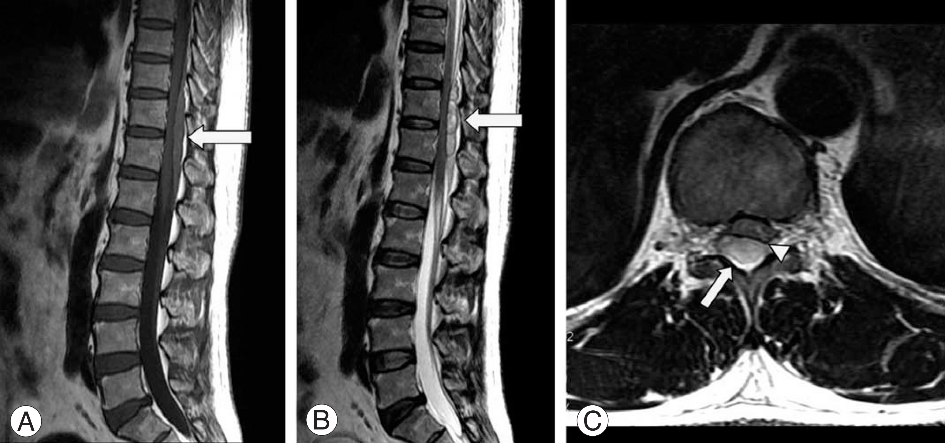
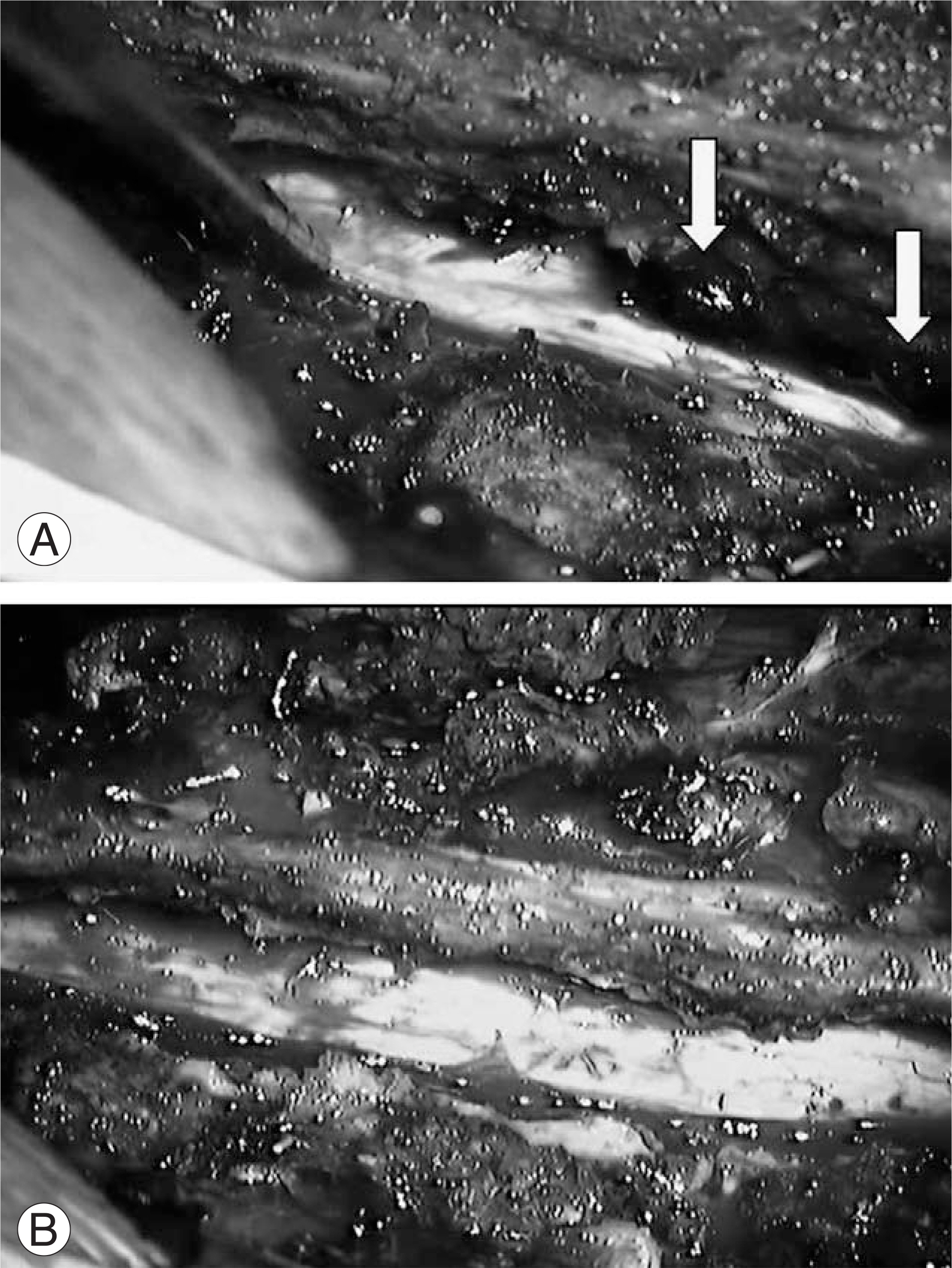
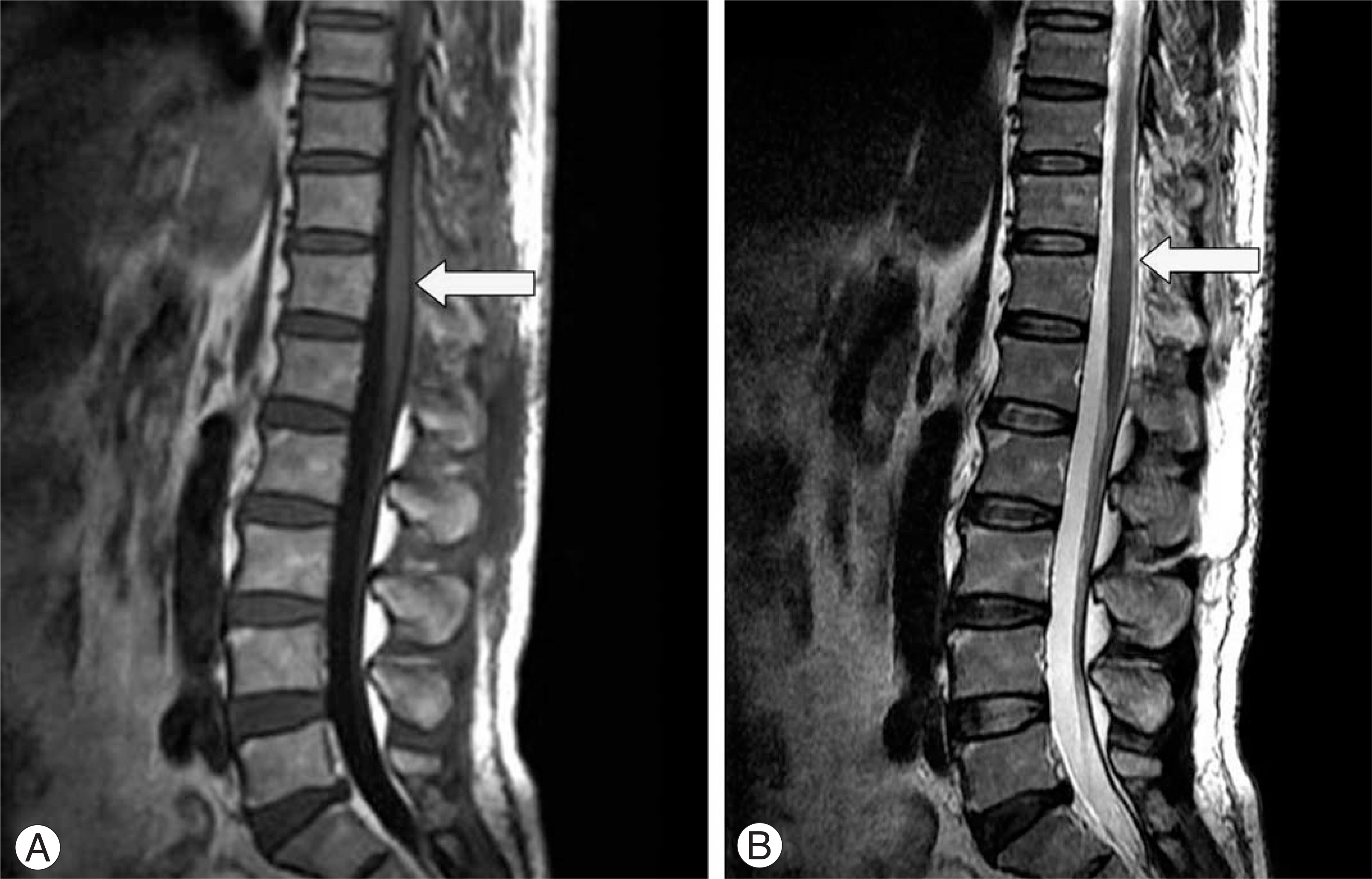
- Spontaneous spinal epidural hematomas without any risk factors, such as spinal tap, trauma, pregnancy, bleeding diathesis, vascular malformations, hypertension, etc. are relatively rare clinical entities. In addition, the clinical suspicion is quite difficult because there are various clinical symptoms according to the size and location of hematoma. However, the speed of diagnosis and initiation of the appropriate treatment are important because the outcome for patients is usually determined by the location and degree of neurological deficits and the duration of dural compression. We report the diagnosis and treatment of spontaneous spinal epidural hematoma in this case with a review of the relevant literature.
MeSH Terms
Figure
Reference
-
01). Holtas S., Heiling M., Lonntoft M. Spontaneous spinal epidural hematoma: finding at MR imaging and clinical correlation. Radiology. 1996. 199:409–413.02). Jackson R. Case of spinal apoplexy. Lancet. 1869. 2:5–6.
Article03). Chung HI., Yim MB., Byun IS., Kim IH. Spontaneous spinal epidural hematoma. J Korean Neurosurg Soc. 1978. 7:145–150.
Article04). Groen RJ., van Alphen HA. Operative treatment of spontaneous spinal epidural hematomas: a study of the factors determining postoperative outcome. Neurosurgery. 1997. 39:494–504.
Article05). Betty RM., Winston KR. Spontaneous cervical epidural hematoma. A consideration of etiology. 1984. 61:143–148.06). Ravid S., Schneider S., Maytal J. Spontaneous spinal epidural hamatoma: an unconnon presentation of a rare disease. Childs Nerv Syst. 2002. 18:345–347.07). Lovblad KO., Baumgartner RW., Zambaz BD., Remon-da L., Ozdoba C., Schroth G. Nontraumatic spinal epidural hematomas. MR features. Acta Radiol. 1997. 38:1–7.08). Wagner S., Forsting M., Hacke W. Spontaneous resolution of a large spinal epidural hematoma: Case report. Neurosurgery. 1996. 38:816–819.
Article09). Duffill J., Sparrow OC., Millar J., Barker CS. Can spontaneous spinal epidural haematoma be managed safely without operation? A report of four cases. J Neurol Neusurg Pychiatry. 2000. 69:816–819.
Article10). Shin JJ., Kuh SU., Cho YE. Surgical management of spontaneous spial epidural hematomas. Eur Spine J. 2006. 15:998–1004.


