Breast Metastasis from Renal Cell Carcinoma: A Case Report
- Affiliations
-
- 1Department of Radiology, Sanggye Paik Hospital, Inje University College of Medicine, Korea. radkimjy@paik.ac.kr
- 2Department of Pathology, Sanggye Paik Hospital, Inje University College of Medicine, Korea.
- 3Department of Surgery, Sanggye Paik Hospital, Inje University College of Medicine, Korea.
- KMID: 2208999
- DOI: http://doi.org/10.3348/jksr.2010.62.1.77
Abstract
- Metastatic breast cancer from renal cell carcinoma is extremely rare and has non-specific findings that include a well circumscribed lesion without calcification on mammography and a well circumscribed hypoechoic lesion without posterior acoustic shadowing on sonography. We report a case of metastatic breast cancer from renal cell carcinoma and describe the radiologic findings in a 63-year-old woman who has no history of primary neoplasm.
MeSH Terms
Figure
-
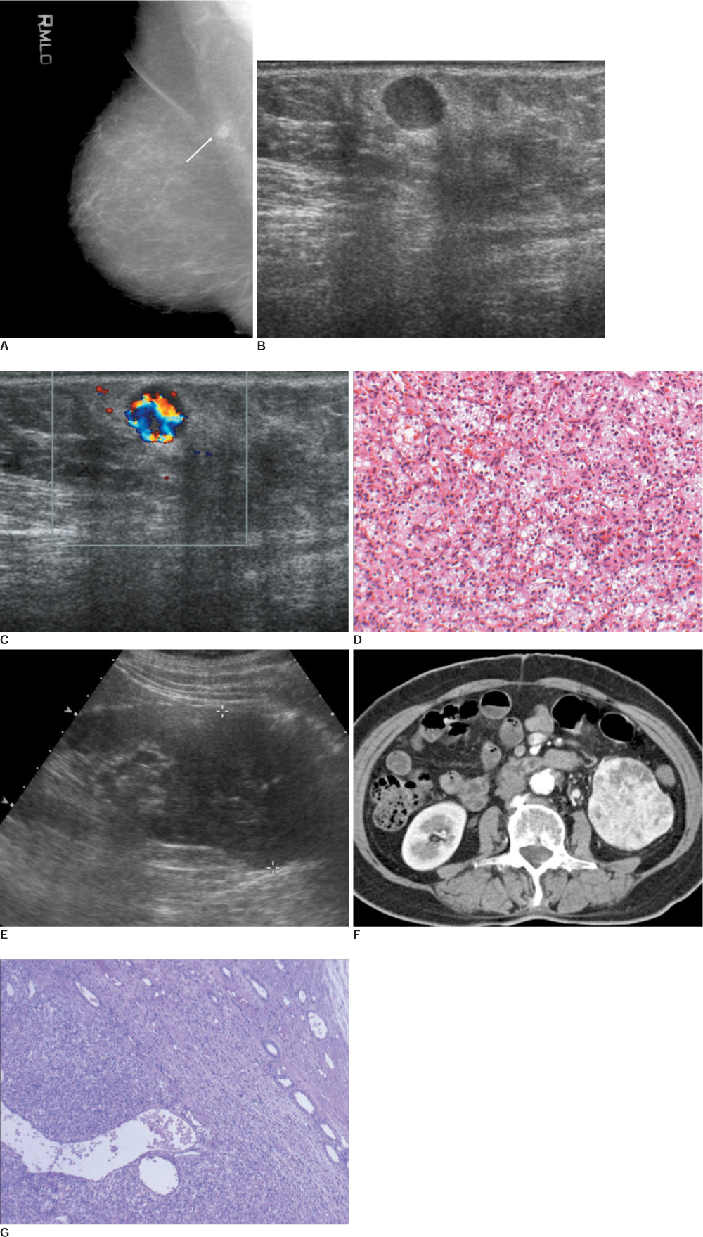
Fig. 1 A 63-year-old woman with metastatic breast cancer from renal cell carcinoma. A. Mediolateral oblique mammography shows a partially circumscribed, partially obscured nodular density in posterior aspect of right breast upper portion (arrow). B. Sonography shows a round shape, well-defined, hypoechoic nodule with slightly thick echogenic halo in subcutaneous layer of right outer breast (arrow). C. Color Doppler examination shows prominent internal vascularity in nodule. D. The tumor cells are nested by the attenuated endothelial cells, showing sinusoidal pattern (H & E, ×100). E. Abdominal sonography shows 7 cm low echoic mass in the left kidney. F. CT of arterial phase shows heterogeneous enhancing mass in left kidney. G. The tumor is relatively demarcated from the normal renal tubules. The tumor reveals sinusoidal vessel pattern. The tumor cells are round to oval with clear cytoplasm and round nuclei, and relatively prominent nucleoli (Fuhrman's Nuclear grade-3 out of 4) (×100).
Reference
-
1. Sandison AT. Metastatic tumors in the breast. Br J Surg. 1959; 47:54–58.2. Hajdu SI, Urban JA. Cancers metastatic to the breast. Cancer. 1972; 29:1691–1696.3. Paulus DD, Libshitz HI. Metastasis to the breast. Radiol Clin North Am. 1982; 20:561–568.4. Toombs BD, Kalisher L. Metastatic disease to the breast: clinical, pathologic, and radiographic features. AJR Am J Roentgenol. 1977; 129:673–676.5. Derchi LE, Rizzatto G, Giuseppetti GM, Dini G, Garaventa A. Metastatic tumors in the breast: sonographic findings. J Ultrasound Med. 1985; 4:69–74.6. Vergier B, Trojani M, de Mascarel I, Coindre JM, Le Treut A. Metastases to the breast: differential diagnosis from primary breast carcinoma. J Surg Oncol. 1991; 48:112–116.7. Lee WK, Cawson JN, Hill PA, Hoang J, Rouse H. Renal cell carcinoma metastasis to the breast: mammographic, sonographic, CT, and pathologic correlation. Breast J. 2007; 13:316–317.8. McLauglin SA, Thiel DD, Smith SL, Wehle MJ, Menke DM. Solitary breast mass as initial presentation of clinically silent metastatic renal cell carcinoma. Breast. 2006; 15:427–429.9. Alzaraa A, Vodovnik A, Montgomery H, Saeed M, Sharma N. Breast metastasis from a renal cell cancer. World J Surg Oncol. 2007; 5:25.10. Stavros AT. Breast ultrasound. Philadelphia: Lippincott Williams & Wilkins;2004. p. 667–670.
- Full Text Links
-
- Actions
-
Cited
- CITED
-
- Close
- Share
-
- Similar articles
-
- Thyroid Metastasis in Pyramidal Lobe from Renal Cell Carcinoma: A Case Report
- A Case of Metastatic Renal Cell Carcinoma to the Gallbladder
- A Case of Subcutaneous Tissue Metastasis of Renal Cell Carcinoma
- Two Cases of Renal Cell Carcinoma Metastatic to the Thyroid Gland
- A Case of Renal Metastasis from a 1-cm Squamous Cell Carcinoma of the Lung Masquerading as Renal Cell Carcinoma
