Multiple Synchronous Central Giant Cell Granulomas of the Maxillofacial Region: A Case Report
- Affiliations
-
- 1Department of Radiology, Pusan National University Hospital, College of Medicine, Pusan National University. Korea. hakjink@pusan.ac.kr
- KMID: 2208988
- DOI: http://doi.org/10.3348/jksr.2010.62.1.11
Abstract
- Multifocal central giant cell granulomas (CGCG) in the maxillofacial region are suggestive of systemic disease such as hyperparathyroidism or an inherited syndrome such as Noonan-like multiple giant cell lesion syndrome. Only 5 cases of multifocal CGCGs in the maxillofacial region without any concomitant systemic disease have currently been reported. We report here on an unusual case of 17-year-old man who presented with multifocal CGCGs of the bilateral posterior mandible and right maxilla and he was without any concomitant systemic disease.
MeSH Terms
Figure
-

Fig. 1 A. The coronal CT scan with a bone window setting shows a relatively well defined, heterogeneous isodense mass (arrows) with diffuse mineralized septa (arrowheads) in the right maxilla. B. The axial CT scan with a bone window setting shows expansile remodeling with cortical thinning of the medial and lateral walls of the maxillary antrum (arrowheads). The mass eroded the adjacent bony structures, such as the anterior wall of the sphenoid sinus (black arrow) and the mass extends to the right antrum and the posterior nasal cavity (white arrow). C. The axial CT scan (below the level of B) shows multiple osteolytic lesions (arrows) with cortical expansion, bone thinning and mineralized septa (arrowheads) in the bilateral posterior mandible.

Fig. 2 A-F. The multiple masses (arrows) in the right maxillary sinus and bilateral posterior mandible show low signal intensity on the T1-weighted images (A, B), heterogeneous low to intermediate signal intensity on the T2-weighted images (C, D) and intense enhancement of the masses (E, F) after the administration of contrast agent.
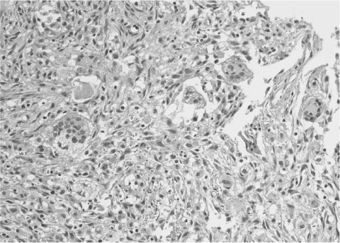
Fig. 3 The biopsy specimen shows several multinucleated giant cells within a background of ovoid and spindle-shaped mesenchymal cells (hematoxylin and eosin staining, magnification×400).
Reference
-
1. Curtis NJ, Walker DM. A case of aggressive multiple metachronous central giant cell granulomas of the jaws: differential diagnosis and management options. Int J Oral Maxillofac Surg. 2005; 34:806–808.2. Murphey MD, Nomikos GC, Flemming DJ, Gannon FH, Temple HT, Kransdorf MJ. Imaging of giant cell tumor and giant cell reparative granuloma of bone: radiologic-pathologic correlation. Radiographics. 2001; 21:1283–1309.3. Edwards PC, Fox J, Fantasia JE, Goldberg J, Kelsch RD. Bilateral central giant cell granulomas of the mandible in an 8-year-old girl with Noonan syndrome (Noonan-like/multiple giant cell lesion syndrome). Oral Surg Oral Med Oral Pathol Oral Radiol Endod. 2005; 99:334–340.4. Junquera LM, Lupi E, Lombardia E, Fresno MF. Multiple and synchronodus peripheral giant cell granulomas of the gums. Ann Otol Rhinol Laryngol. 2002; 111:751–753.5. Martins WD, de Oliveira Ribas M, Braosi AP, Machado MA, Lima AA. Multiple giant cell lesions of the maxillofacial skeleton. J Oral Maxillofac Surg. 2007; 65:1250–1253.6. Morris JM, Lane JI, Witte RJ, Thompson DM. Giant cell reparative granuloma of the nasal cavity. AJNR Am J Neuroradiol. 2004; 25:1263–1265.7. Smith PG, Marrogi AJ, Delfino JJ. Multifocal central giant cell lesions of the maxillofacial skeleton: a case report. J Oral Maxillofac Surg. 1990; 48:300–305.8. Cakirer S. Quiz case. Giant cell reparative granuloma of the maxillary sinus. Eur J Radiol. 2002; 44:24–27.
- Full Text Links
-
- Actions
-
Cited
- CITED
-
- Close
- Share
-
- Similar articles
-
- Central giant cell granuloma in mandible: report of a case
- Central giant cell lesion of the mandible in a 2-year old girl
- Central giant cell granuloma and cementifying fibroma occurring in the same lesion of right mandibular body: a case report
- Synchronous thyroid carcinoma and squamous cell carcinoma: A case report
- A Case of a Central Giant Cell Granuloma in the Right Zygomatic Bone
