Radiologic Findings of Foreign Body Granuloma by the Bee Sting: A Case Report
- Affiliations
-
- 1Department of Radiology, Kangnam Sacred Heart Hospital, College of Medicine, Hallym University, Seoul, Korea. ikyang@hallym.or.kr
- 2Departments of Pathology, Hallym University College of Medicine, Kangnam Sacred Heart Hospital, Seoul, Korea.
- KMID: 2208925
- DOI: http://doi.org/10.3348/jksr.2010.62.3.283
Abstract
- Bee sting therapy is a folk remedy used for arthralgia. An adverse reaction to bee sting therapy can be variable, ranging from a local inflammatory reaction to generalized anaphylaxis. There have been reports of dermatologic findings pertaining to bee sting granulomas, which results from a foreign body reaction to the persistence of venom and stinger at the sting site. However to the best of our knowledge, the radiologic findings of bee sting granulomas have not been reported on in Korea. We describe the ultrasound and MRI findings of bee sting granulomas at the lower extremity in a 36-year-old woman who underwent bee-sting therapy for osteoarthritis of the knee joints 3 months prior.
MeSH Terms
Figure
-
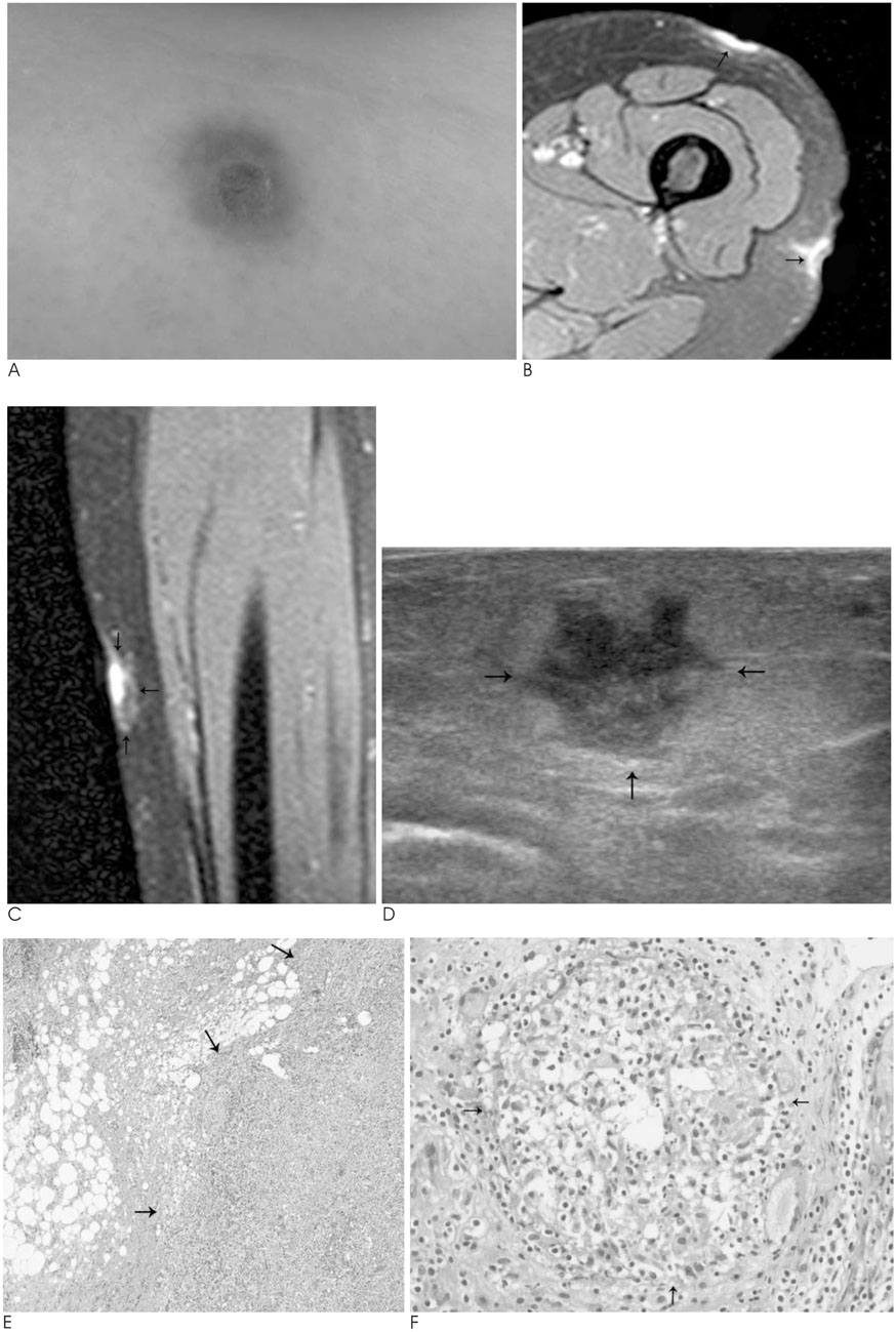
Fig. 1 Bee sting granuloma of the left thigh in a 36-year-old woman. A. Photograph of the bee sting granuloma in left thigh is seen as erythematous patch with ulcer. B, C. Fat suppressed proton density MR images show high signal intensity lesions with edema (arrows) in the subcutaneous fat layer of left thigh. D. High resolution ultrasonography shows irregular, lobulated hypoechoic lesion (arrows) with surrounding hyperechoic fat infiltration in the subcutaneous layer of left thigh. E. Photomicrography shows a large aggregation of inflammatory cells with histiocytes and lymphocytes (arrows). Destruction of fat lobule, degeneration of fat cells with formation of lymphoid follicles are also noted (H & E stain, ×40). F. Well-formed nonnecrotizing granuloma (arrows), an aggregation of epithelioid histiocytes surrounded by lymphocytes is shown (H & E stain, ×200).
Reference
-
1. Park JH, Kim JG, Cha SH, Park SD. Eosinophilic foreign body granuloma after multiple self-administered bee stings. Br J Dermatol. 1998; 139:1102–1105.2. Yu HJ, Lee CW, Yang HY, Kim JS, Kim YS. Three Cases of Bee-sting Granuloma. Korean J Dermatol. 1998; 36:914–917.3. Lee SH, Sung KJ, Koh JK. Foreign-body granuloma after honeybee acupuncture. Ann Dermatol. 1996; 8:215–217.4. Lee CW, Cho JH, YU HJ, Yang HY, Park CK, Park MH. Bee-sting granulomas in the skin. Dermatology. 1996; 193:355–356.5. Hur W, Ahn SK, Lee SH, Kang WH. Cutaneous reaction induced by retained bee stinger. J Dermatol. 1991; 18:736–739.6. Ando A, Hatori M, Hagiwara Y, Isefuku S, Itoi E. Imaging features of foreign body granuloma in the lower extremities mimicking a soft tissue neoplasm. UPS J Med Sci. 2009; 114:46–51.7. Nakamura T, Kusuzaki K, Matsubara T, Matsumine A, Uchida A. Foreign-body granulomas in the trunk and extremities may stinulate malignant soft-tissue tumors: report of three cases. Acta Radiol. 2008; 49:80–83.
- Full Text Links
-
- Actions
-
Cited
- CITED
-
- Close
- Share
-
- Similar articles
-
- Foreign-body Granuloma After Honeybee Acupuncture
- A Case of Foreign Body Granuloma with Skin Necrosis Occurring after Bee Sting Therapy
- Foreign Body Granuloma Following Dried Honey Bee Venom (Apitoxin Inj) Injection
- Three Cases of Bee-sting Granuloma
- A Case of Foreign Body Granuloma after Squalene Injection by Non-dermatologists
