Multiorgan Involvements of Cowden Disease in a 50-Year-Old Woman: A Case Report and Literature Overview
- Affiliations
-
- 1Department of Radiology, St. Vincent's Hospital, The Catholic University of Korea College of Medicine, Suwon, Korea. radiodoc@catholic.ac.kr
- KMID: 2208809
- DOI: http://doi.org/10.3348/jksr.2013.69.3.251
Abstract
- Cowden disease is the prototype of phosphate and, tensin homologue deleted on the chromosome 10 (PTEN) hamartoma tumor syndrome caused by germline mutations in the tumor suppressor PTEN, which is characterized by multiple developmentally disorganized benign growths, hamartomas, with an increased risk of both benign and malignant tumors. We present another case of Cowden disease in a 50-year-old woman. Besides the diagnostic criteria of Cowden disease, she had various manifestations in thyroid, lung, spleen, liver, pancreas, and muscle. As far as we know, it is the first case showing radiographic findings of hamartomatous lesions in thyroid, spleen, and pancreas, associated with Cowden disease.
MeSH Terms
Figure
-

Fig. 1 Mammography shows a spiculated irregular mass with pleomorphic microcalcifications at the upper portion of the right central breast (arrow). Ipsilateral lymphadenopathy is associated.

Fig. 2 Chest CT scans with mediastinal window setting. A. A small discrete nodule is noted in the periphery of the lung (arrow). The nodule do not show FDG uptake on PET-CT scan (not shown). On the follow-up CT scan, there was no interval change of the nodule for 6 months (not shown). B. A small lung cyst (arrowhead) is also seen with an unclear relation to CD, but it may be a component of the hamartomatous lesions. Note.-CD = Cowden disease, FDG = fluorodeoxyglucose, PET-CT = positron emission tomography-CT

Fig. 3 CT scan shows diffuse enlargement of the thyroid gland with macrocalcifications. Note the multiple fat containing nodules (arrowheads), suggesting thyrolipomas.

Fig. 4 Contrast enhanced CT scans of abdomen. A. Hemangioma-like mass with fat density portion is seen in the right hepatic lobe (arrow). B. Multiple tiny fat density nodules (arrowheads) noted in pancreas tail suggest lipomas. The aforementioned hepatic mass is also visible (arrow).The lesions are noted with no FDG uptake on PET-CT (not shown). C. Low-density polyp is suspected within the sigmoid colon (arrow). The lesion is confirmed by colonoscopy. D. Fat containing enhanced nodule is noted within the right gluteus muscle (arrow). Note.-FDG = fluorodeoxyglucose, PET-CT = positron emission tomography-CT
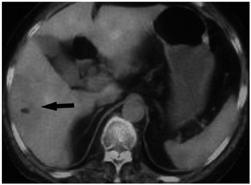
Fig. 5 PET-CT shows no FDG uptake in the hepatic lesion (arrow). Note.-FDG = fluorodeoxyglucose, PET-CT = positron emission tomography-CT
Reference
-
1. Hobert JA, Eng C. PTEN hamartoma tumor syndrome: an overview. Genet Med. 2009; 11:687–694.2. Rademaker J, Kim YJ, Leibecke T, Raman SS, Voit C. Cowden disease: CT findings in three patients. Abdom Imaging. 2005; 30:204–207.3. Criteira TNCsT. NCCN guidelines Version 1. 2011 Cowden syndrome. 2011. Available from: www.nccn.org.4. Farooq A, Walker LJ, Bowling J, Audisio RA. Cowden syndrome. Cancer Treat Rev. 2010; 36:577–583.5. Pilarski R. Cowden syndrome: a critical review of the clinical literature. J Genet Couns. 2009; 18:13–27.6. Laury AR, Bongiovanni M, Tille JC, Kozakewich H, Nosé V. Thyroid pathology in PTEN-hamartoma tumor syndrome: characteristic findings of a distinct entity. Thyroid. 2011; 21:135–144.
- Full Text Links
-
- Actions
-
Cited
- CITED
-
- Close
- Share
-
- Similar articles
-
- Breast Cancer in Cowden Syndrome: A Case Report
- A case of Cowden's syndrome associated with Lhermitte-Duclos disease
- A Case of Cowden's Disease Associated with Breast Cancer
- Candida Esophagitis in a Patient with Cowden's Syndrome: A Case Report
- Cowden's disease--a report on the first case in Korea and literature review
