Detection of Hepatocellular Carcinoma: Comparison of Gadoxetic Acid-Enhanced MRI, Diffusion-Weighted Imaging, and Combined Interpretation at 3 T MRI
- Affiliations
-
- 1Department of Radiology and Research Institute of Radiological Science, Severance Hospital, Yonsei University College of Medicine, Seoul, Korea. gafield2@yuhs.ac
- 2Department of Radiology, Hallym University College of Medicine, Seoul, Korea.
- KMID: 2208804
- DOI: http://doi.org/10.3348/jksr.2013.69.3.213
Abstract
- PURPOSE
To compare diffusion-weighted imaging (DWI) and gadoxetic acid-enhanced (Gdx) magnetic resonance imaging (MRI), whether alone or in combination, for the detection of hepatocellular carcinoma (HCC) by using 3 T.
MATERIALS AND METHODS
84 HCCs in 66 patients (57 men, 9 women; mean age 69.2 years) were examined using 3 T MRI. DWI (b values 0, 50, and 800 sec/mm2) and dynamic gadoxetic acid-enhanced MRI as well as hepatobiliary phase were performed. Images were retrospectively reviewed by two radiologists to compare the diagnostic performances of DWI and Gdx MRI alone and in combination for the detection of HCC. Alternative free response receiver operating characteristic analysis and comparison of sensitivities were used for statistical analysis.
RESULTS
The sensitivity of Gdx set (73/84, 87%) was significantly higher than that of DWI set (60.5/84, 72%) for both observers. The Az values of DWI and Gdx MRI for the detection of HCC were not statistically significant for either observer (Az for DWI = 0.818 and 0.864, Az for Gdx MR = 0.902 and 0.842, respectively, p = 0.107 for observer 1 and p = 0.738 for observer 2). The combination of both techniques did not increase the sensitivities of detecting HCC in either observer. When lesions smaller than 2 cm were considered, the DWI set yielded a significantly lower sensitivity as compared with either the Gdx set alone or the combination set.
CONCLUSION
Gadoxetic acid-enhanced MRI was better than DWI for detection the HCC by using 3 T MRI. The combination of DWI and Gdx MRI did not contribute to the successful detection of HCC.
MeSH Terms
Figure
-
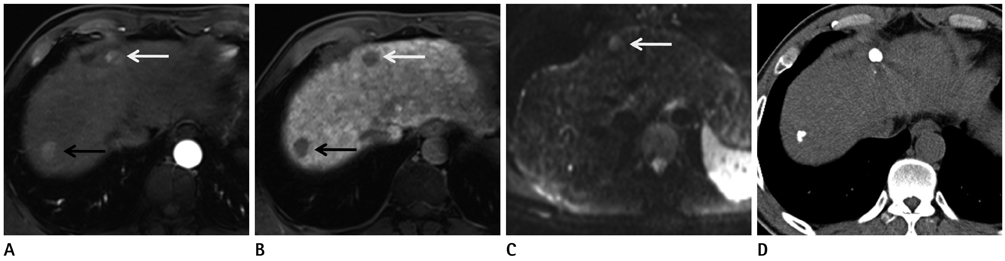
Fig. 1 Two hepatocellular carcinomas in a 48-year-old man. A. Gadoxetic acid-enhanced MRI image during the arterial phase shows two enhancing masses, one in the right hepatic lobe (black arrow) and one in segment 4 (white arrow). B. A hepatobiliary phase image 20 minutes after contrast administration shows hypointense masses at the same locations (black and white arrows). C. Diffusion-weighted image (DWI) demonstrates a hyperintense lesion (arrow) at S4. However, the lesion in the right hepatic lobe is not depicted on DWI. D. Follow-up unenhanced CT one month after transarterial chemoembolization shows iodized oil retention in S4 and the right lobe.
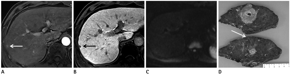
Fig. 2 Hepatocellular carcinoma in a 42-year-old man. The main mass, 4 cm sized expansile hypervascular tumor at S6, is not shown here. A, B. Gadoxetic acid-enhanced MR image during arterial phase (A) and hepatobiliary phase (B) show a satellite nodule in the right hepatic lobe (arrows). C. The lesions are not visible on DWI using b values of 50 s/mm2. D. Photograph of the surgical specimen shows a main mass and a small hepatocellular carcinoma (arrow). Note.-DWI = diffusion-weighted imaging
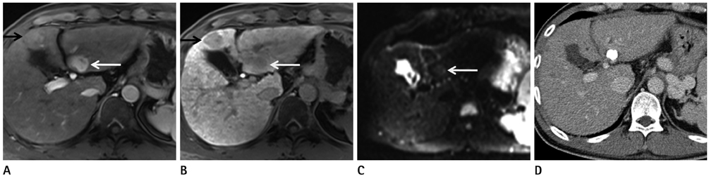
Fig. 3 Hepatocellular carcinoma in a 45-year-old man. A. Gadoxetic acid-enhanced MR images during the arterial phase show two enhancing nodules in the left hepatic lobe (black and white arrows). B. The lesions show hypointensity on hepatobiliary phase images at the same location (black and white arrows). C. Only the lesion at S3 is visible on DWI using b values of 800 s/mm2 (arrow). D. Follow-up CT shows compact iodized oil retention at S3. The lesion at S4 did not show any enhancement or washout during the follow-up period of one and a half years. Note.-DWI = diffusion-weighted imaging
Reference
-
1. El-Serag HB. Hepatocellular carcinoma: an epidemiologic view. J Clin Gastroenterol. 2002; 35:5 Suppl 2. S72–S78.2. El-Serag HB. Hepatocellular carcinoma: recent trends in the United States. Gastroenterology. 2004; 127:5 Suppl 1. S27–S34.3. Baron RL, Brancatelli G. Computed tomographic imaging of hepatocellular carcinoma. Gastroenterology. 2004; 127:5 Suppl 1. S133–S143.4. Kim SH, Kim SH, Lee J, Kim MJ, Jeon YH, Park Y, et al. Gadoxetic acid-enhanced MRI versus triple-phase MDCT for the preoperative detection of hepatocellular carcinoma. AJR Am J Roentgenol. 2009; 192:1675–1681.5. Kwak HS, Lee JM, Kim YK, Lee YH, Kim CS. Detection of hepatocellular carcinoma: comparison of ferumoxides-enhanced and gadolinium-enhanced dynamic three-dimensional volume interpolated breath-hold MR imaging. Eur Radiol. 2005; 15:140–147.6. Kanematsu M, Kondo H, Goshima S, Tsuge Y, Watanabe H. Magnetic resonance imaging of hepatocellular carcinoma. Oncology. 2008; 75:Suppl 1. 65–71.7. Kim MJ, Choi JY, Chung YE, Choi SY. Magnetic resonance imaging of hepatocellular carcinoma using contrast media. Oncology. 2008; 75:Suppl 1. 72–82.8. Willatt JM, Hussain HK, Adusumilli S, Marrero JA. MR Imaging of hepatocellular carcinoma in the cirrhotic liver: challenges and controversies. Radiology. 2008; 247:311–330.9. Bartolozzi C, Crocetti L, Lencioni R, Cioni D, Della Pina C, Campani D. Biliary and reticuloendothelial impairment in hepatocarcinogenesis: the diagnostic role of tissue-specific MR contrast media. Eur Radiol. 2007; 17:2519–2530.10. Frericks BB, Loddenkemper C, Huppertz A, Valdeig S, Stroux A, Seja M, et al. Qualitative and quantitative evaluation of hepatocellular carcinoma and cirrhotic liver enhancement using Gd-EOB-DTPA. AJR Am J Roentgenol. 2009; 193:1053–1060.11. Hammerstingl R, Huppertz A, Breuer J, Balzer T, Blakeborough A, Carter R, et al. Diagnostic efficacy of gadoxetic acid (Primovist)-enhanced MRI and spiral CT for a therapeutic strategy: comparison with intraoperative and histopathologic findings in focal liver lesions. Eur Radiol. 2008; 18:457–467.12. Huppertz A, Haraida S, Kraus A, Zech CJ, Scheidler J, Breuer J, et al. Enhancement of focal liver lesions at gadoxetic acid-enhanced MR imaging: correlation with histopathologic findings and spiral CT--initial observations. Radiology. 2005; 234:468–478.13. Reimer P, Rummeny EJ, Shamsi K, Balzer T, Daldrup HE, Tombach B, et al. Phase II clinical evaluation of Gd-EOB-DTPA: dose, safety aspects, and pulse sequence. Radiology. 1996; 199:177–183.14. Ahn SS, Kim MJ, Lim JS, Hong HS, Chung YE, Choi JY. Added value of gadoxetic acid-enhanced hepatobiliary phase MR imaging in the diagnosis of hepatocellular carcinoma. Radiology. 2010; 255:459–466.15. Nasu K, Kuroki Y, Nawano S, Kuroki S, Tsukamoto T, Yamamoto S, et al. Hepatic metastases: diffusion-weighted sensitivity-encoding versus SPIO-enhanced MR imaging. Radiology. 2006; 239:122–130.16. Hollingsworth KG, Lomas DJ. Influence of perfusion on hepatic MR diffusion measurement. NMR Biomed. 2006; 19:231–235.17. Koh DM, Collins DJ. Diffusion-weighted MRI in the body: applications and challenges in oncology. AJR Am J Roentgenol. 2007; 188:1622–1635.18. Taouli B, Koh DM. Diffusion-weighted MR imaging of the liver. Radiology. 2010; 254:47–66.19. Choi JY, Kim MJ, Chung YE, Kim JY, Jones AC, de Becker J, et al. Abdominal applications of 3.0-T MR imaging: comparative review versus a 1.5-T system. Radiographics. 2008; 28:e30.20. Merkle EM, Dale BM, Paulson EK. Abdominal MR imaging at 3T. Magn Reson Imaging Clin N Am. 2006; 14:17–26.21. Soher BJ, Dale BM, Merkle EM. A review of MR physics: 3T versus 1.5T. Magn Reson Imaging Clin N Am. 2007; 15:277–290. v22. Choi SH, Lee JM, Yu NC, Suh KS, Jang JJ, Kim SH, et al. Hepatocellular carcinoma in liver transplantation candidates: detection with gadobenate dimeglumine-enhanced MRI. AJR Am J Roentgenol. 2008; 191:529–536.23. Piana G, Trinquart L, Meskine N, Barrau V, Beers BV, Vilgrain V. New MR imaging criteria with a diffusion-weighted sequence for the diagnosis of hepatocellular carcinoma in chronic liver diseases. J Hepatol. 2011; 55:126–132.24. Namimoto T, Yamashita Y, Sumi S, Tang Y, Takahashi M. Focal liver masses: characterization with diffusion-weighted echo-planar MR imaging. Radiology. 1997; 204:739–744.25. Parikh T, Drew SJ, Lee VS, Wong S, Hecht EM, Babb JS, et al. Focal liver lesion detection and characterization with diffusion-weighted MR imaging: comparison with standard breath-hold T2-weighted imaging. Radiology. 2008; 246:812–822.26. Zech CJ, Herrmann KA, Dietrich O, Horger W, Reiser MF, Schoenberg SO. Black-blood diffusion-weighted EPI acquisition of the liver with parallel imaging: comparison with a standard T2-weighted sequence for detection of focal liver lesions. Invest Radiol. 2008; 43:261–266.27. Nasu K, Kuroki Y, Tsukamoto T, Nakajima H, Mori K, Minami M. Diffusion-weighted imaging of surgically resected hepatocellular carcinoma: imaging characteristics and relationship among signal intensity, apparent diffusion coefficient, and histopathologic grade. AJR Am J Roentgenol. 2009; 193:438–444.28. Shinya S, Sasaki T, Nakagawa Y, Guiquing Z, Yamamoto F, Yamashita Y. The efficacy of diffusion-weighted imaging for the detection of colorectal cancer. Hepatogastroenterology. 2009; 56:128–132.29. Xu PJ, Yan FH, Wang JH, Lin J, Ji Y. Added value of breath-hold diffusion-weighted MRI in detection of small hepatocellular carcinoma lesions compared with dynamic contrast-enhanced MRI alone using receiver operating characteristic curve analysis. J Magn Reson Imaging. 2009; 29:341–349.30. Holzapfel K, Bruegel M, Eiber M, Ganter C, Schuster T, Heinrich P, et al. Characterization of small (≤10 mm) focal liver lesions: value of respiratory-triggered echo-planar diffusion-weighted MR imaging. Eur J Radiol. 2010; 76:89–95.31. Nishie A, Tajima T, Ishigami K, Ushijima Y, Okamoto D, Hirakawa M, et al. Detection of hepatocellular carcinoma (HCC) using super paramagnetic iron oxide (SPIO)-enhanced MRI: Added value of diffusion-weighted imaging (DWI). J Magn Reson Imaging. 2010; 31:373–382.32. Hardie AD, Naik M, Hecht EM, Chandarana H, Mannelli L, Babb JS, et al. Diagnosis of liver metastases: value of diffusion-weighted MRI compared with gadolinium-enhanced MRI. Eur Radiol. 2010; 20:1431–1441.33. Le Bihan D, Breton E, Lallemand D, Aubin ML, Vignaud J, Laval-Jeantet M. Separation of diffusion and perfusion in intravoxel incoherent motion MR imaging. Radiology. 1988; 168:497–505.34. Goshima S, Kanematsu M, Kondo H, Yokoyama R, Tsuge Y, Shiratori Y, et al. Evaluating local hepatocellular carcinoma recurrence post-transcatheter arterial chemoembolization: is diffusion-weighted MRI reliable as an indicator? J Magn Reson Imaging. 2008; 27:834–839.35. Kim YK, Kim CS, Han YM, Lee YH. Detection of liver malignancy with gadoxetic acid-enhanced MRI: is addition of diffusion-weighted MRI beneficial? Clin Radiol. 2011; 66:489–496.36. Choi JY, Choi JS, Kim MJ, Lim JS, Park MS, Kim JH, et al. Detection of hepatic hypovascular metastases: 3D gradient echo MRI using a hepatobiliary contrast agent. J Magn Reson Imaging. 2010; 31:571–578.37. Kim YK, Ko SW, Hwang SB, Kim CS, Yu HC. Detection and characterization of liver metastases: 16-slice multidetector computed tomography versus superparamagnetic iron oxide-enhanced magnetic resonance imaging. Eur Radiol. 2006; 16:1337–1345.38. Kim YK, Lee JM, Kim CS. Gadobenate dimeglumine-enhanced liver MR imaging: value of dynamic and delayed imaging for the characterization and detection of focal liver lesions. Eur Radiol. 2004; 14:5–13.39. Wang H, Wang XY, Jiang XX, Ye ZX. Comparison of diffusion-weighted with T2-weighted Imaging for detection of small hepatocellular carcinoma in cirrhosis: preliminary quantitative study at 3-T. Acad Radiol. 2010; 17:239–243.
- Full Text Links
-
- Actions
-
Cited
- CITED
-
- Close
- Share
-
- Similar articles
-
- Diagnosis of Hepatocellular Carcinoma with Gadoxetic Acid-Enhanced MRI: 2016 Consensus Recommendations of the Korean Society of Abdominal Radiology
- Gadoxetic acid-enhanced magnetic resonance imaging: Hepatocellular carcinoma and mimickers
- Pathology-MRI Correlation of Hepatocarcinogenesis: Recent Update
- Gadoxetic Acid (Gd-EOB-DTPA)-Enhanced MRI versus Gadobenate Dimeglumine (Gd-BOPTA)-Enhanced MRI for Preoperatively Detecting Hepatocellular Carcinoma: an Initial Experience
- Hepatic Angiomyolipoma Presenting as a Hyperintense Lesion During the Hepatobiliary Phase of Gadoxetic Acid Enhanced-MRI: a Case Report
