Added Value of Magnetic Resonance Imaging in Staging of Malignant Pleural Mesothelioma
- Affiliations
-
- 1Department of Radiology and Research Institute of Radiology, University of Ulsan College of Medicine, Asan Medical Center, Korea. ejinchae@gmail.com
- KMID: 2206922
- DOI: http://doi.org/10.13104/jksmrm.2013.17.3.232
Abstract
- PURPOSE
We investigated the possible added value of magnetic resonance imaging (MR) in staging of malignant pleural mesothelioma (MPM) compared to computed tomography (CT).
MATERIALS AND METHODS
We retrospectively enrolled 20 patients (M;F = 14:6; mean age, 53.5 yrs) who diagnosed as MPM by histology and underwent CT and MR at initial evaluation from Jan 1997 to Dec 2012. Two radiologists performed clinical staging by using CT alone or MR alone in consensus. In patients underwent surgery (n = 13), we evaluated the diagnostic accuracy of CT and MR in terms of staging compared to surgical staging. In all patients, we compared clinical staging of CT only and CT with MR.
RESULTS
The diagnostic accuracy for T staging of CT only was 23.1% (3/13) and that of combined CT and MR was 38.5% (5/13), respectively. Among 13 patients underwent surgery, surgical stage was higher than combined CT and MR stage in 5 patients, but lower in 3 patients. CT only and combined CT and MR agreed in 85.0% (17/20). In cases of disagree (15.0%, 3/20), combined CT and MR showed higher stage than CT only.
CONCLUSION
Combined CT and MR increases the diagnostic accuracy in staging of MPM compared to CT only and is important in determining the appropriate treatment in patients being considered for surgery.
MeSH Terms
Figure
-

Fig. 1 Differences between clinical T stage and corrected surgical T stage in patients with available surgical stage.

Fig. 2 A representative case of upstaging by MR compared to surgical stage. a. A CT image shows circumferential pleural thickening involving visceral and parietal pleura. Areas of obliteration of extrapleural fat probably over the expected line of endothoracic fascia suggest chest wall invasion (T4) (arrows). b. Gadolinium-enhanced T1-weighted MR image shows some indentation of chest wall by the pleural mass suggesting chest wall invasion (T4) (arrows). However, patient underwent pleuropneumonectomy and surgical and histologic findings revealed no evidence of chest wall invasion (T2).
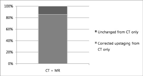
Fig. 3 Differences between clinical stage by CT only and CT and MR.

Fig. 4 A representative case of upstaging by MR compared to CT. a. A CT image shows pleural effusion with diffuse pleural thickening. Soft tissue density lesions are suspicious in intercostal spaces in the lower portion of hemithorax, however chest wall invasion is not clear. b. Gadolinium-enhanced, 3-dimensional, gradient recalled echo sequence MR image clearly shows enhancing soft tissue lesions in the intercostal spaces suggesting diffuse chest wall invasion (T4) (arrow).
Reference
-
1. Jung KW, Won YJ, Kong HJ, Oh CM, Seo HG, Lee JS. Cancer statistics in Korea: incidence, mortality, survival and prevalence in 2010. Cancer Res Treat. 2013; 45:1–14.2. Teta MJ, Mink PJ, Lau E, Sceurman BK, Foster ED. US mesothelioma patterns 1973-2002: indicators of change and insights into background rates. Eur J Cancer Prev. 2008; 17:525–534.3. Price B, Ware A. Mesothelioma trends in the United States: an update based on Surveillance, Epidemiology, and End Results Program data for 1973 through 2003. Am J Epidemiol. 2004; 159:107–112.4. Delgermaa V, Takahashi K, Park EK, Le GV, Hara T, Sorahan T. Global mesothelioma deaths reported to the World Health Organization between 1994 and 2008. Bull World Health Organ. 2011; 89:716–724.5. Cao C, Tian DH, Pataky KA, Yan TD. Systematic review of pleurectomy in the treatment of malignant pleural mesothelioma. Lung Cancer. 2013; 81:319–327.6. Erasmus JJ, Truong MT, Smythe WR, et al. Integrated computed tomography-positron emission tomography in patients with potentially resectable malignant pleural mesothelioma: Staging implications. J Thorac Cardiovasc Surg. 2005; 129:1364–1370.7. Scherpereel A, Astoul P, Baas P, et al. Guidelines of the European Respiratory Society and the European Society of Thoracic Surgeons for the management of malignant pleural mesothelioma. Eur Respir J. 2010; 35:479–495.8. Martino D, Pass HI. Integration of multimodality approaches in the management of malignant pleural mesothelioma. Clin Lung Cancer. 2004; 5:290–298.9. Neumann V, Loseke S, Nowak D, Herth FJ, Tannapfel A. Malignant pleural mesothelioma: incidence, etiology, diagnosis, treatment, and occupational health. Dtsch Arztebl Int. 2013; 110:319–326.10. Sugarbaker DJ, Flores RM, Jaklitsch MT, et al. Resection margins, extrapleural nodal status, and cell type determine postoperative long-term survival in trimodality therapy of malignant pleural mesothelioma: results in 183 patients. J Thorac Cardiovasc Surg. 1999; 117:54–63.11. Rusch V, Baldini EH, Bueno R, et al. The role of surgical cytoreduction in the treatment of malignant pleural mesothelioma: meeting summary of the International Mesothelioma Interest Group Congress, September 11-14, 2012, Boston, Mass. J Thorac Cardiovasc Surg. 2013; 145:909–910.12. Nowak AK. CT, RECIST, and malignant pleural mesothelioma. Lung Cancer. 2005; 49:Suppl 1. S37–S40.13. Patz EF Jr, Shaffer K, Piwnica-Worms DR, et al. Malignant pleural mesothelioma: value of CT and MR imaging in predicting resectability. AJR Am J Roentgenol. 1992; 159:961–966.14. Heelan RT, Rusch VW, Begg CB, Panicek DM, Caravelli JF, Eisen C. Staging of malignant pleural mesothelioma: comparison of CT and MR imaging. AJR Am J Roentgenol. 1999; 172:1039–1047.15. Wang ZJ, Reddy GP, Gotway MB, et al. Malignant pleural mesothelioma: evaluation with CT, MR imaging, and PET. Radiographics. 2004; 24:105–119.16. Boiselle PM, Patz EF Jr, Vining DJ, Weissleder R, Shepard JA, McLoud TC. Imaging of mediastinal lymph nodes: CT, MR, and FDG PET. Radiographics. 1998; 18:1061–1069.17. Karabulut N, Martin DR, Yang M, Tallaksen RJ. MR imaging of the chest using a contrast-enhanced breath-hold modified three-dimensional gradient-echo technique: comparison with two-dimensional gradient-echo technique and multidetector CT. AJR Am J Roentgenol. 2002; 179:1225–1233.18. Hintze C, Dinkel J, Biederer J, Heussel CP, Puderbach M. New procedures. Comprehensive staging of lung cancer by MRI. Radiologe. 2010; 50:699–705.19. Kajiwara N, Akata S, Uchida O, et al. Cine MRI enables better therapeutic planning than CT in cases of possible lung cancer chest wall invasion. Lung Cancer. 2010; 69:203–208.20. Plathow C, Staab A, Schmaehl A, et al. Computed tomography, positron emission tomography, positron emission tomography/computed tomography, and magnetic resonance imaging for staging of limited pleural mesothelioma: initial results. Invest Radiol. 2008; 43:737–744.21. Zahid I, Sharif S, Routledge T, Scarci M. What is the best way to diagnose and stage malignant pleural mesothelioma. Interact Cardiovasc Thorac Surg. 2011; 12:254–259.22. Rusch VW. A proposed new international TNM staging system for malignant pleural mesothelioma. From the International Mesothelioma Interest Group. Chest. 1995; 108:1122–1128.23. Treasure T, Lang-Lazdunski L, Waller D, et al. Extra-pleural pneumonectomy versus no extra-pleural pneumonectomy for patients with malignant pleural mesothelioma: clinical outcomes of the Mesothelioma and Radical Surgery (MARS) randomised feasibility study. Lancet Oncol. 2011; 12:763–772.24. Datta A, Smith R, Fiorentino F, Treasure T. Surgery in the treatment of malignant pleural mesothelioma: recruitment into trials should be the default position. Thorax. 2013; Epub.25. Gill RR. Imaging of mesothelioma. Recent Results Cancer Res. 2011; 189:27–43.26. Gill RR, Gerbaudo VH, Jacobson FL, et al. MR imaging of benign and malignant pleural disease. Magn Reson Imaging Clin N Am. 2008; 16:319–339.27. Donmez FY, Yekeler E, Saeidi V, Tunaci A, Tunaci M, Acunas G. Dynamic contrast enhancement patterns of solitary pulmonary nodules on 3D gradient-recalled echo MRI. AJR Am J Roentgenol. 2007; 189:1380–1386.28. Lee VS, Lavelle MT, Krinsky GA, Rofsky NM. Volumetric MR imaging of the liver and applications. Magn Reson Imaging Clin N Am. 2001; 9:697–716.29. Weber U, Lambert RG, Rufibach K, et al. Anterior chest wall inflammation by whole-body magnetic resonance imaging in patients with spondyloarthritis: lack of association between clinical and imaging findings in a cross-sectional study. Arthritis Res Ther. 2012; 14:R3.
- Full Text Links
-
- Actions
-
Cited
- CITED
-
- Close
- Share
-
- Similar articles
-
- Malignant Mesothelioma Causing Bloody Pleural Effusion
- A Case of Malignant Mesothelioma with Pleural Effusion
- Malignant Mesothelioma Presenting as a Giant Chest, Abdominal and Pelvic Wall Mass
- Added values of transrectal ultrasonography to magnetic resonance imaging in characterizing prostate cancer: A narrative review
- A Case of Malignant Pericardial Mesothelioma with Atypical CT and MR Imaging Pattern
