Background Parenchymal Enhancement on Breast MRI in Breast Cancer Patients : Impact on Biopsy Rate and Cancer Yield
- Affiliations
-
- 1Department of Radiology, Seoul St. Mary's Hospital, College of Medicine, The Catholic University of Korea, Korea. rad-ksh@catholic.ac.kr
- KMID: 2206921
- DOI: http://doi.org/10.13104/jksmrm.2013.17.3.224
Abstract
- PURPOSE
To evaluate the potential effects of background parenchymal enhancement of MR imaging in diagnosed breast cancer patients on the rate of additional biopsy and resultant cancer yield.
MATERIALS AND METHODS
322 patients who were diagnosed with breast cancer and had undergone breast MR imaging were included in this study. Two radiologists reviewed the MRI for degree of background parenchymal enhancement and additional suspicious lesions described as BI-RADS category 4 or 5 on radiologic reports. Biopsy was done for these lesions, pathology reports were reviewed to calculate the cancer yield.
RESULTS
Background parenchymal enhancement of MR imaging in a total of 322 patients were classified as minimal degree 47.5%, mild degree 28.9%, moderate degree 12.4% and marked degree 11.2%. Among these 332 patients, MR imaging of 70 patients showed additional suspicious malignant lesions described as BI-RADS category 4 or 5, and consequently, 66 patients underwent biopsy. Biopsy rates in those with minimal or mild background parenchymal enhancement and those with moderate and marked background parenchymal enhancement were 19.9% and 22.3% (p-value 0.77) respectively. Cancer yields in those with minimal or mild background parenchymal enhancement and those with moderate and marked background parenchymal enhancement were 6.5% and 5.2% (p value 0.88) respectively. Both these results did not show stastically significant difference between the two groups.
CONCLUSION
The degree of background parenchymal enhancement in MR imaging of breast cancer patients did not significantly impact additional biopsy rates or cancer yields.
Figure
-
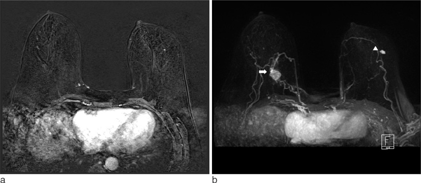
Fig. 1 A 57-year-old woman with right breast cancer and minimal parenchymal enhancement. a. Axial early T1-weighted contrast-enhanced MR subtraction image shows minimal parenchymal enhancement. b. On Maximum-intensity-projection (MIP) image, there is index cancer in the right breast (arrow). Another lobular, homogeneously enhancing mass is seen in the left breast (arrow head), which was classified as BI-RADS category C4A, low suspicion of malignancy, and proved to be fibrocystic disease (FCD) on US-guided biopsy.
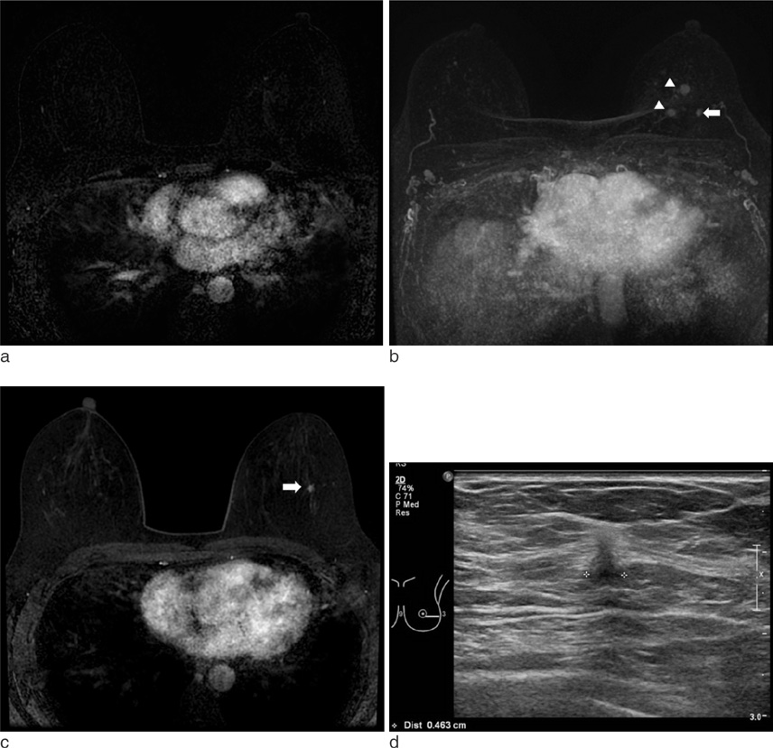
Fig. 2 A 46-year-old woman with left breast cancer and minimal parenchymal enhancement. a. Axial early T1-weighted contrast-enhanced MR subtraction image shows minimal parenchymal enhancement. b. On MIP image, two pathologic proven invasive ductal carcinomas are noted at the 10-11 o'clock direction of left breast (arrow heads). Another enhancing mass is seen at the 3 o'clock direction of left breast (arrow). c. Axial T1-weighted contrast-enhanced MRI with fat supression at early dynamic phase shows this mass to be an enhancing, oval, spiculated mass in the left breast (arrow), consistent with BI-RADS category C4C, moderate suspicion of malignancy. d. An irregular, spiculated, non-parallel-oriented mass is detected on 2nd look US, which corresponds to the lesion on MRI and later proved to be invasive ductal carcinoma on US-guided biopsy.
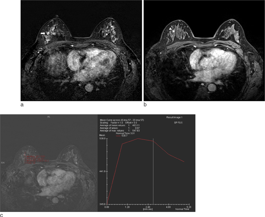
Fig. 3 A 46-year-old woman with right breast cancer and marked parenchymal enhancement. a. Axial early T1-weighted contrast-enhanced MR subtraction image shows marked parenchymal enhancement and an index cancer (arrow), known invasive lobular carcinoma, in the right breast. b. Axial T1-weighted contrast-enhanced MRI with fat supression at early dynamic phase shows another oval, homogeneously enhancing mass at the 11 o'clock direction of right breast (arrow head), which was classified as BI-RADS category C4A, low suspicion of malignancy. c. Kinetic curve of the mass at the 11 o'clock direction shows delayed washout pattern; the mass proved to be lobular carcinoma in situ (LCIS) on US-guided biopsy.
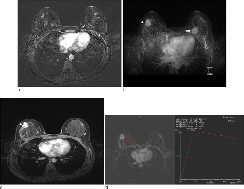
Fig. 4 A 40-year-old woman with left breast cancer and moderate parenchymal enhancement. a. Axial early T1-weighted contrast-enhanced MR subtraction image shows moderate parenchymal enhancement. b. MIP image shows moderate parenchymal enhancement and an index cancer, known invasive ductal carcinoma, in the left breast (arrow). Another enhancing mass similar to the index cancer is seen in the right breast (arrow head). c. On axial T1-weighted contrast-enhanced MRI with fat supression at early dynamic phase, an lobular, heterogeneously enhancing mass is seen in the right breast (arrow head), which was categorized as BI-RADS C4C, moderate suspicion of malignancy, d. The kinetic curve of the right breast mass shows delayed washout pattern. The mass proved to be tubular adenoma on US-guided biopsy.
Reference
-
1. Kuhl CK, Bieling HB, Gieseke J, et al. Healthy premenopausal breast parenchyma in dynamic contrast-enhanced MR imaging of the breast: normal contrast medium enhancement and cyclical-phase dependency. Radiology. 1997; 203:137–144.2. Delille JP, Slanetz PJ, Yeh ED, Kopans DB, Halpern EF, Garrido L. Hormone replacement therapy in postmenopausal women: breast tissue perfusion determined with MR imaging--initial observations. Radiology. 2005; 235:36–41.3. DeMartini WB, Liu F, Peacock S, Eby PR, Gutierrez RL, Lehman CD. Background parenchymal enhancement on breast MRI: impact on diagnostic performance. AJR Am J Roentgenol. 2012; 198:W373–W380.4. Hambly NM, Liberman L, Dershaw DD, Brennan S, Morris EA. Background parenchymal enhancement on baseline screening breast MRI: impact on biopsy rate and short-interval follow-up. AJR Am J Roentgenol. 2011; 196:218–224.5. Morris EA. Diagnostic breast MR imaging: current status and future directions. Magn Reson Imaging Clin N Am. 2010; 18:57–74.6. Uematsu T, Yuen S, Kasami M, Uchida Y. Comparison of magnetic resonance imaging, multidetector row computed tomography, ultrasonography, and mammography for tumor extension of breast cancer. Breast Cancer Res Treat. 2008; 112:461–474.7. Uematsu T, Kasami M, Watanabe J. Background enhancement of mammary glandular tissue on breast dynamic MRI: imaging features and effect on assessment of breast cancer extent. Breast Cancer. 2012; 19:259–265.8. Kuhl C. The Current status of breast MR imaging Part I. Choice of technique, image interpretation, diagnostic accuracy, and transfer to clinical practice. Radiology. 2007; 244:356–378.9. Uematsu T, Kasami M, Watanabe J. Does the degree of background enhancement in breast MRI affect the detection and staging of breast cancer? Eur Radiol. 2011; 21:2261–2267.
- Full Text Links
-
- Actions
-
Cited
- CITED
-
- Close
- Share
-
- Similar articles
-
- Breast Cancer Screening with MRI
- The Study of Relation between Breast parenchymal Patterns and Breast Cancer by Mammography
- Usefulness of Preoperative Breast MRI in Breast Cancer Diagnosed After Excisional Biopsy
- Effect of the Menstrual Cycle on Background Parenchymal Enhancement Observed on Breast MRIs in Korean Women
- The Highligts of 28th Annual Meeting of San Antonio Breast Cancer Symposium
