J Korean Soc Radiol.
2015 Sep;73(3):158-163. 10.3348/jksr.2015.73.3.158.
Effect of the Menstrual Cycle on Background Parenchymal Enhancement Observed on Breast MRIs in Korean Women
- Affiliations
-
- 1Department of Radiology and Research Institute of Radiological Science, Severance Hospital, Yonsei University College of Medicine, Seoul, Korea. mines@yuhs.ac
- KMID: 2043605
- DOI: http://doi.org/10.3348/jksr.2015.73.3.158
Abstract
- PURPOSE
To evaluate the effect of the menstrual cycle on background parenchymal enhancement observed on breast MRIs in Korean women, and to suggest an optimal period for scheduling breast MRIs.
MATERIALS AND METHODS
Between March and December 2012, 214 premenopausal breast cancer patients who underwent breast MRIs for preoperative evaluation were included. Levels of background parenchymal enhancement were retrospectively compared according to the menstrual cycle.
RESULTS
There was no significant difference between levels of background parenchymal enhancement (minimal, mild, moderate, and marked) according to the weeks of the menstrual cycle. However, the 1st and 2nd week of the menstrual cycle showed a significantly higher proportion of patients with minimal background parenchymal enhancement than the 3rd and 4th week of the menstrual cycle (47.0% vs. 32.0%; p = 0.025).
CONCLUSION
For screening purposes and for the follow-up of Korean breast cancer patients, breast MRIs should be performed during the 1st or 2nd week of the menstrual cycle.
MeSH Terms
Figure
-
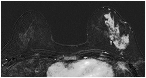
Fig. 1 A 42-year-old woman with left breast cancer who underwent a preoperative breast MRI during the second week of her menstrual cycle. The first subtraction axial image shows minimal background parenchymal enhancement at her right breast.

Fig. 2 A 34-year-old women with left breast cancer who underwent a preoperative breast MRI during the third week of her menstrual cycle. The first subtraction axial image shows marked background parenchymal enhancement at her right breast.
Reference
-
1. Saslow D, Boetes C, Burke W, Harms S, Leach MO, Lehman CD, et al. American Cancer Society guidelines for breast screening with MRI as an adjunct to mammography. CA Cancer J Clin. 2007; 57:75–89.2. Palestrant S, Comstock CE, Moy L. Approach to breast magnetic resonance imaging interpretation. Radiol Clin North Am. 2014; 52:563–583.3. DeMartini WB, Liu F, Peacock S, Eby PR, Gutierrez RL, Lehman CD. Background parenchymal enhancement on breast MRI: impact on diagnostic performance. AJR Am J Roentgenol. 2012; 198:W373–W380.4. Kuhl CK, Bieling HB, Gieseke J, Kreft BP, Sommer T, Lutterbey G, et al. Healthy premenopausal breast parenchyma in dynamic contrast-enhanced MR imaging of the breast: normal contrast medium enhancement and cyclical-phase dependency. Radiology. 1997; 203:137–144.5. Morris EA, Comstock C, Lee C, Lehman CD, Ikeda DM, Newstead GM, et al. ACR BI-RADS® Magnetic Resonance Imaging. In : . Breast Imaging Reporting and Data System. Reston, VA: American College of Radiology;2013.6. Mann RM, Balleyguier C, Baltzer PA, Bick U, Colin C, Cornford E, et al. Breast MRI: EUSOBI recommendations for womens information. Eur Radiol. 2015; 05. 23. [Epub]. DOI: 10.1007/s00330-015-3807-z.7. Gweon HM, Cho N, Han W, Yi A, Moon HG, Noh DY, et al. Breast MR imaging screening in women with a history of breast conservation therapy. Radiology. 2014; 272:366–373.8. Müller-Schimpfle M, Ohmenhaüser K, Stoll P, Dietz K, Claussen CD. Menstrual cycle and age: influence on parenchymal contrast medium enhancement in MR imaging of the breast. Radiology. 1997; 203:145–149.9. Vogel PM, Georgiade NG, Fetter BF, Vogel FS, McCarty KS Jr. The correlation of histologic changes in the human breast with the menstrual cycle. Am J Pathol. 1981; 104:23–34.10. Delille JP, Slanetz PJ, Yeh ED, Kopans DB, Garrido L. Physiologic changes in breast magnetic resonance imaging during the menstrual cycle: perfusion imaging, signal enhancement, and influence of the T1 relaxation time of breast tissue. Breast J. 2005; 11:236–241.11. Harlow SD, Ephross SA. Epidemiology of menstruation and its relevance to women's health. Epidemiol Rev. 1995; 17:265–286.12. Kajihara M, Goto M, Hirayama Y, Okunishi S, Kaoku S, Konishi E, et al. Effect of the menstrual cycle on background parenchymal enhancement in breast MR imaging. Magn Reson Med Sci. 2013; 12:39–45.13. Kang SS, Ko EY, Han BK, Shin JH, Hahn SY, Ko ES. Background parenchymal enhancement on breast MRI: influence of menstrual cycle and breast composition. J Magn Reson Imaging. 2014; 39:526–534.14. Fowler PA, Casey CE, Cameron GG, Foster MA, Knight CH. Cyclic changes in composition and volume of the breast during the menstrual cycle, measured by magnetic resonance imaging. Br J Obstet Gynaecol. 1990; 97:595–602.15. Graham SJ, Stanchev PL, Lloyd-Smith JO, Bronskill MJ, Plewes DB. Changes in fibroglandular volume and water content of breast tissue during the menstrual cycle observed by MR imaging at 1.5 T. J Magn Reson Imaging. 1995; 5:695–670.
- Full Text Links
-
- Actions
-
Cited
- CITED
-
- Close
- Share
-
- Similar articles
-
- Menstruation and Sleep
- Mammographic Changes in Breast Cancer Patients Treated with Tamoxifen
- Background Parenchymal Enhancement on Breast MRI in Breast Cancer Patients : Impact on Biopsy Rate and Cancer Yield
- Metabolic Activity of Normal Glandular Tissue on ¹â¸F-Fluorodeoxyglucose Positron Emission Tomography/Computed Tomography: Correlation with Menstrual Cycles and Parenchymal Enhancements
- Influence of the Phase of Menstrual Cycle on Postoperative Nausea and Vomiting after Breast Cancer Surgery
