Solitary Capillary Hemangioma of the Lung: A Report of Two Cases
- Affiliations
-
- 1Department of Pathology, Samsung Medical Center, Sungkyunkwan University School of Medicine, Seoul, Korea. Joungho.han@samsung.net
- 2Department of Thoracic Surgery, Samsung Medical Center, Sungkyunkwan University School of Medicine, Seoul, Korea.
- 3Department of Radiology, Samsung Medical Center, Sungkyunkwan University School of Medicine, Seoul, Korea.
- KMID: 2199838
- DOI: http://doi.org/10.6058/jlc.2012.11.2.102
Abstract
- It has long been recognized that tumors of capillary vessels in the lung are extremely rare. While pulmonary capillary hemangiomatosis, which is a rare cause of pulmonary hypertension, is relatively well known, there have been only a few reports of solitary capillary hemangioma (SCH) of the lung. Herein we report two cases of SCH. Both cases were first detected as a solitary nodule of the lung in chest computed tomography images. Both lesions were recognized as early lung cancer and surgical resections were performed. At low power view, one was not encapsulated but was well delineated from the non-neoplastic lung and the other was pseudo-capsulated. Both tumors consisted of uniform capillaries with cuboidal or flattened endothelial cells. There was no cytologic atypia. Endothelial cells were highlighted by CD31 stain. Awareness of this entity is important for pathologists for differential diagnosis of a solitary pulmonary nodule.
Keyword
MeSH Terms
Figure
-
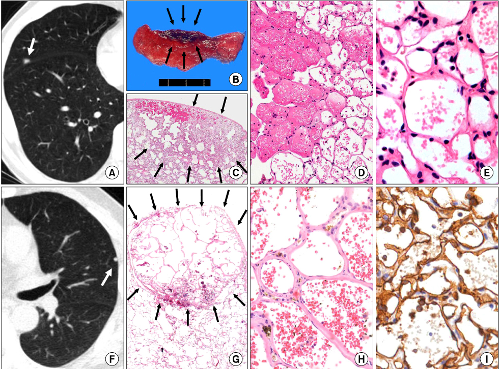
Fig. 1 (A) In the first case, chest computed tomography (CT) shows a small single nodular mass (arrow). (B) Wedge resected lung shows an ill-defined soft hemorrhagic mass (arrows). (C) The tumor is not encapsulated but well delineated from the non-neoplastic lung (arrows). (D, E) The tumor consists of uniform capillaries, with cuboidal or flattened endothelial cells. There is no cellular atypia or mitosis. (F) In the second case, chest CT shows a single nodular mass (arrow). (G) The tumor is round in shape and is pseudo-encapsulated (arrows). (H) The tumor consists of dilated capillaries. There is no cellular atypia or mitosis. (I) CD31 immunohistochemical stain highlights endothelial tumor cells (1:80, Dako, Glostrup, Denmark) (C and G, H&E, ×10; D, H&E, ×100; E and H, H&E, ×200; I, CD31, ×200).
Reference
-
1. Umezu H, Naito M, Yagisawa K, Hattori A, Aizawa Y. An autopsy case of pulmonary capillary hemangiomatosis without evidence of pulmonary hypertension. Virchows Arch. 2001. 439:586–592.
Article2. Wagenvoort CA, Beetstra A, Spijker J. Capillary haemangiomatosis of the lungs. Histopathology. 1978. 2:401–406.
Article3. Abrahams NA, Colby TV, Pearl RH, Chipps BE, Juris AL, Leslie KO. Pulmonary hemangiomas of infancy and childhood: report of two cases and review of the literature. Pediatr Dev Pathol. 2002. 5:283–292.
Article4. Bowyer JJ, Sheppard M. Capillary haemangioma presenting as a lung pseudocyst. Arch Dis Child. 1990. 65:1162–1164.
Article5. Fugo K, Matsuno Y, Okamoto K, et al. Solitary capillary hemangioma of the lung: report of 2 resected cases detected by high-resolution CT. Am J Surg Pathol. 2006. 30:750–753.6. Galliani CA, Beatty JF, Grosfeld JL. Cavernous hemangioma of the lung in an infant. Pediatr Pathol. 1992. 12:105–111.
Article7. Kim GY, Kim J, Choi YS, Kim HJ, Ahn G, Han J. Sixteen cases of sclerosing hemangioma of the lung including unusual presentations. J Korean Med Sci. 2004. 19:352–358.
Article
- Full Text Links
-
- Actions
-
Cited
- CITED
-
- Close
- Share
-
- Similar articles
-
- Capillary Hemangioma of Testis
- Cutis Marmorata Telangiectatica Congenita: A Rare Clinical Manifestation of Capillary Hemangioma?
- Episcleral Capillary Hemangioma
- A Case Report of Multiple Capillary Hemangioma in a Chronic Myeloid Leukemia Patient Taking Tyrosine Kinase Inhibitors
- Capillary Hemangioma of the Right Ventricle: A Case Report
