J Korean Acad Prosthodont.
2011 Apr;49(2):161-167. 10.4047/jkap.2011.49.2.161.
Influence of porcelain re-firing on the formation of surface bubble and on the change in shade of metal-ceramic crown exposed to artificial saliva
- Affiliations
-
- 1Department of Prosthodontics, School of Dentistry, Seoul National University, Seoul, Korea. jhoyang@snu.ac.kr
- KMID: 2196123
- DOI: http://doi.org/10.4047/jkap.2011.49.2.161
Abstract
- PURPOSE
The purpose of this study was to evaluate the influence of porcelain re-firing on the formation of surface bubble and on the change in shade of metal-ceramic crown exposed to artificial saliva.
MATERIALS AND METHODS
Thirty disk-shaped specimens were made in 10 mm diameter with 0.5 mm metal core thickness and 1 mm ceramic thickness. A spectroradiometer was used to determine the CIE Lab coordinates. The number and size of surface bubble were observed with a stereomicroscope. After the exposure to artificial saliva for 7 days, re-firing was performed at glazing temperature. After re-firing, the CIE Lab were calculated, and the number and size of surface bubble were observed again. The change in shade was expressed with DeltaE. Statistical analysis was done with paired t-test for the change in the number of surface bubble and student t-test for the change in the size of surface bubble (alpha= 0.05).
RESULTS
Shade difference was calculated 2.14 DeltaE units. The mean number of surface bubble was 1.33 +/- 1.49 before re-firing, 3.27 +/- 2.90 after re-firing. After re-firing, the number of surface bubble was significantly increased (P<.05). The mean size of surface bubble was 81.97 +/- 32.03 microm before re-firing, 142.94 +/- 47.40 microm after re-firing. After re-firing, the size of surface bubble was significantly increased (P<.05).
CONCLUSION
Shade change after re-firing was perceptible (DeltaE < 2.0) and clinically acceptable (DeltaE < 3.7). The number and size of surface bubble was significantly increased after re-firing. Further investigation to decrease the surface bubble on the extra oral repair of metal-ceramic crown, will be needed in future study.
Keyword
MeSH Terms
Figure
-
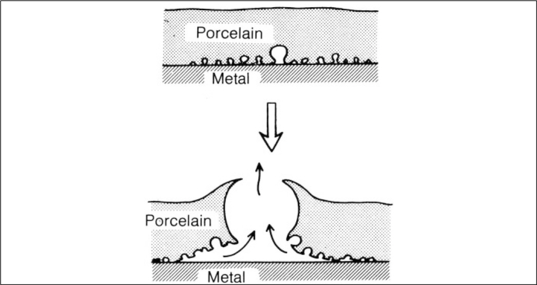
Fig. 1. Generated large bubble.
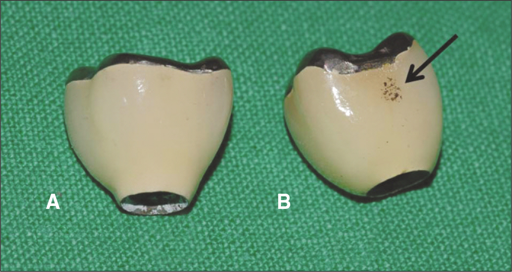
Fig. 2. Effect of immersion in acetone. A: Removal of organic materials, B: Combustion of organic materials.
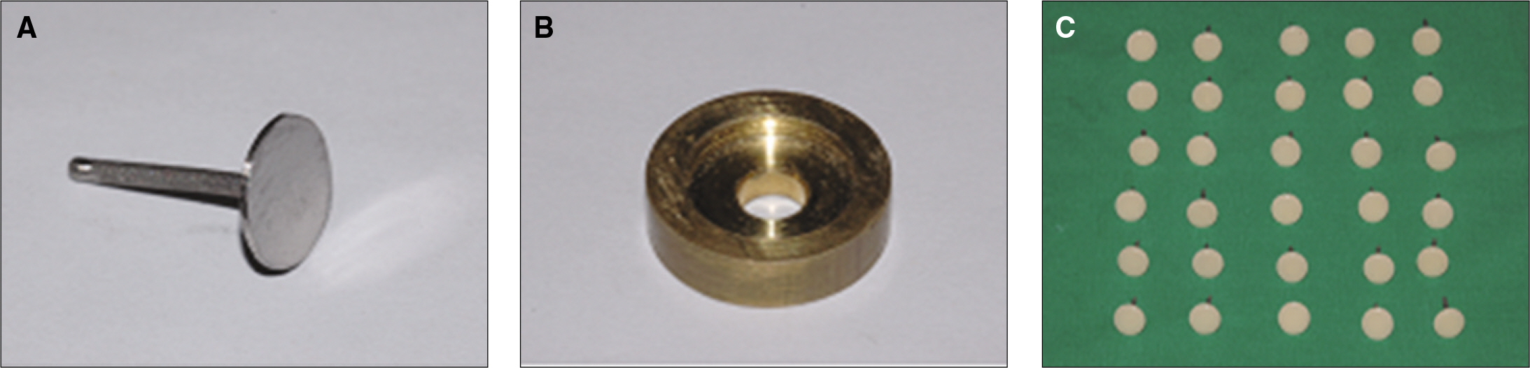
Fig. 3. Procedure of specimen fabrication. A: Metal coping, B: Mold for porcelain build up, C: fabricated specimens.
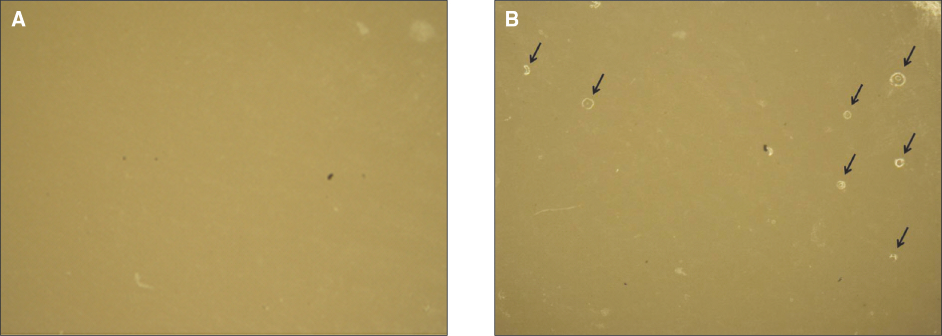
Fig. 4. Increase of air bubbles on porcelain surface after re-firing was detected in the stereomicroscope images. (arrows) (Original magnification, ×20). A: Before re-firing, B: After re-firing.
Reference
-
1.Yamamoto M. Metal-Ceramics. Chicago: Quintessence;Publishing Co.1985. p. 119–34.2.Pjetursson BE., Bra¨gger U., Lang NP., Zwahlen M. Comparison of survival and complication rates of tooth-supported fixed dental prostheses (FDPs) and implant-supported FDPs and single crowns (SCs). Clin Oral Implants Res. 2007. 18:97–113.
Article3.Galiatsatos AA. An indirect repair technique for fractured metal-ceramic restorations: a clinical report. J Prosthet Dent. 2005. 93:321–3.
Article4.Kelly JR., Nishimura I., Campbell SD. Ceramics in dentistry: historical roots and current perspectives. J Prosthet Dent. 1996. 75:18–32.
Article5.Stannard JG., Marks L., Kanchanatawewat K. Effect of multiple firing on the bond strength of selected matched porcelain-fused-to-metal combinations. J Prosthet Dent. 1990. 63:627–9.
Article6.Mackert JR Jr., Williams AL. Microcracks in dental porcelain and their behavior during multiple firing. J Dent Res. 1996. 75:1484–90.
Article7.Isgro` G., Kleverlaan CJ., Wang H., Feilzer AJ. The influence of multiple firing on thermal contraction of ceramic materials used for the fabrication of layered all-ceramic dental restorations. Dent Mater. 2005. 21:557–64.8.Ritter JE. Predicting lifetimes of materials and material structures. Dent Mater. 1995. 11:142–6.
Article9.Calamia JR. Etched porcelain veneers: the current state of the art. Quintessence Int. 1985. 16:5–12.10.Clyde JS., Gilmour A. Porcelain veneers: a preliminary review. Br Dent J. 1988. 164:9–14.
Article11.Nicholls JI. Tensile bond of resin cements to porcelain veneers. J Prosthet Dent. 1988. 60:443–7.
Article12.Aboush YE. Removing saliva contamination from porcelain veneers before bonding. J Prosthet Dent. 1998. 80:649–53.
Article13.Phark JH., Duarte S Jr., Kahn H., Blatz MB., Sadan A. Influence of contamination and cleaning on bond strength to modified zirconia. Dent Mater. 2009. 25:1541–50.
Article14.Yang B., Wolfart S., Scharnberg M., Ludwig K., Adelung R., Kern M. Influence of contamination on zirconia ceramic bonding. J Dent Res. 2007. 86:749–53.
Article15.Barghi N., Goldberg . Porcelain shade stability after repeated firing. J Prosthet Dent. 1977. 37:173–5.
Article16.Barghi N., Richardson JT. A study of various factors influencing shade of bonded porcelain. J Prosthet Dent. 1978. 39:282–4.
Article17.Jorgenson MW., Goodkind RJ. Spectrophotometric study of five porcelain shades relative to the dimensions of color, porcelain thickness, and repeated firings. J Prosthet Dent. 1979. 42:96–105.
Article18.Seghi RR., Hewlett ER., Kim J. Visual and instrumental colorimetric assessments of small color differences on translucent dental porcelain. J Dent Res. 1989. 68:1760–4.
Article19.Johnston WM., Kao EC. Assessment of appearance match by visual observation and clinical colorimetry. J Dent Res. 1989. 68:819–22.
Article20.Douglas RD., Steinhauer TJ., Wee AG. Intraoral determination of the tolerance of dentists for perceptibility and acceptability of shade mismatch. J Prosthet Dent. 2007. 97:200–8.
Article21.Yilmaz B., Ozçelik TB., Wee AG. Effect of repeated firings on the color of opaque porcelain applied on different dental alloys. J Prosthet Dent. 2009. 101:395–404.
Article22.Uludag B., Usumez A., Sahin V., Eser K., Ercoban E. The effect of ceramic thickness and number of firings on the color of ceramic systems: an in vitro study. J Prosthet Dent. 2007. 97:25–31.
Article23.Ozturk O., Uludag B., Usumez A., Sahin V., Celik G. The effect of ceramic thickness and number of firings on the color of two all-ce-ramic systems. J Prosthet Dent. 2008. 100:99–106.
Article24.Jones DW., Wilson HJ. Porosity in dental ceramics. Br Dent J. 1975. 138:16–21.
Article25.Cheung KC., Darvell BW. Sintering of dental porcelain: effect of time and temperature on appearance and porosity. Dent Mater. 2002. 18:163–73.
Article26.Hofstede TM., Ercoli C., Graser GN., Tallents RH., Moss ME., Zero DT. Influence of metal surface finishing on porcelain porosity and beam failure loads at the metal-ceramic interface. J Prosthet Dent. 2000. 84:309–17.
Article27.Anusavice KJ., Phillips RW. Phillips' science of dental materials. 7th ed.New York: Elsevier;2003. p. 672.28.Zhang Y., Griggs JA., Benham AW. Influence of powder/liquid mixing ratio on porosity and translucency of dental porcelains. J Prosthet Dent. 2004. 91:128–35.
Article
- Full Text Links
-
- Actions
-
Cited
- CITED
-
- Close
- Share
-
- Similar articles
-
- A study of the shear bond strength of metal brackets and ceramic brackets and the condition after debonding
- Comparison of fracture strength between hybrid-ceramic crown and metal-ceramic crown
- Evaluation of the marginal and internal gap of metal-ceramic crown fabricated with a selective laser sintering technology: two- and three-dimensional replica techniques
- The effect of repeated firings on the color change and surface roughness of dental ceramics
- Effect of cooling rate during simulated porcelain firing and post-firing heat treatment on the hardness of Pd–Ag–In-Sn metal-ceramic alloy
