3-D Finite element stress analysis in screw-type, cement-type, and combined-type implant fixed partial denture designs
- Affiliations
-
- 1Department of Prosthodontics, Graduate School of Clinical Dental Science, The Catholic University of Korea, Korea. seokgyuk@paran.com
- KMID: 2195585
- DOI: http://doi.org/10.4047/jkap.2009.47.4.365
Abstract
- STATEMENT OF PROBLEMS: Stress analysis on implant components of the combined screw- and cement-retained implant prosthesis has not investigated yet.
PURPOSE: The purpose of this study was to assess the load distribution characteristics of implant prostheses with the different prosthodontic retention types, such as cement-type, screw-type and combined type by using 3-dimensional finite element analysis.
MATERIAL AND METHODS: A 3-dimensional finite element model was created in which two SS II implants (Osstem Co. Ltd.) were placed in the areas of the first premolar and the first molar in the mandible , and three-unit fixed partial dentures with four different retention types were fabricated on the two SS II implants. Model 1 was a cement-retained implant restoration made on two cement-retained type abutments (Comocta abutment; Osstem Co. Ltd.), and Model 2 was a screw-retained implant restoration made on the screw-retained type abutments (Octa abutment; Osstem Co. Ltd.). Model 3 was a combined type implant restoration made on the cement-retained type abutment (Comocta abutment) for the first molar and the screw-retained type abutment (Octa abutment) for the first premolar. Lastly, Model 4 was a combined type implant restoration made on the screw-retained type abutment (Octa abutment) for the first molar and the cement-retained type abutment (Comocta abutment) for the first premolar. Average masticatory force was applied on the central fossa in a vertical direction, and on the buccal cusp in a vertical and oblique direction for each model. Von-Mises stress patterns on alveolar bone, implant body, abutment, abutment screw, and prosthetic screw around implant prostheses were evaluated through 3-dimensional finite element analysis.
RESULTS
Model 2 showed the lowest von Mises stress. In all models, the von Mises stress distribution of cortical bone, cancellous bone and implant body showed the similar pattern. Regardless of loading conditions and type of abutment system, the stress of bone was concentrated on the cortical bone. The von-Mises stress on abutment, abutment screw, and prosthetic screw showed the lower values for the screw-retained type abutment than for the cement-retained type abutment regardless of the model type. There was little reciprocal effect of the abutment system between the molar and the premolar position. For all models, buccal cusp oblique loading caused the largest stress, followed by buccal cusp vertical loading and center vertical loading.
CONCLUSION
Within the limitation of the FEA study, the combined type implant prosthesis did not demonstrate more stress around implant components than the cement type implant prosthesis. Under the assumption of ideal passive fit, the screw-type implant prosthesis showed the least stress around implant components.
MeSH Terms
Figure
-
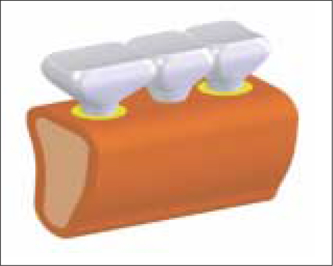
Fig. 1. 3-dimensional experimental model of mandible.
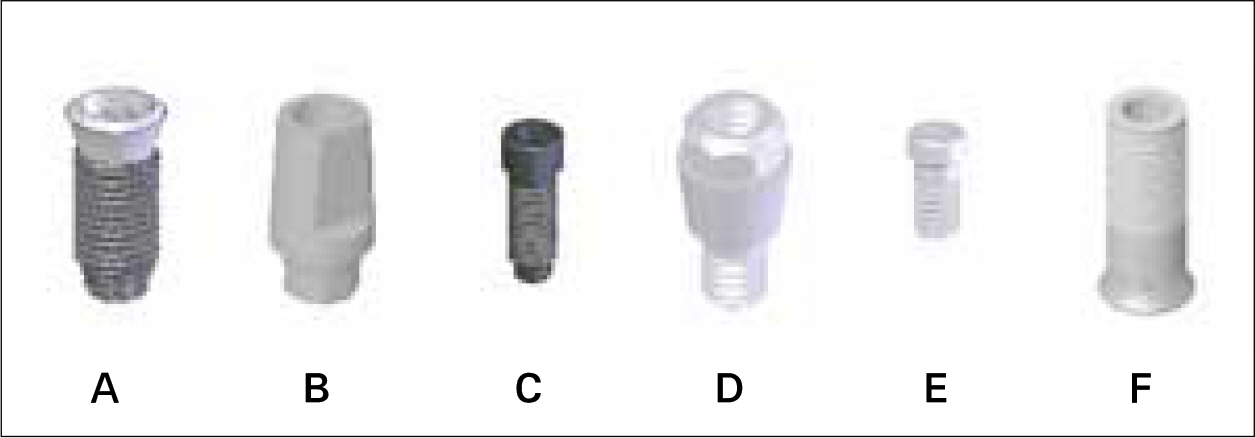
Fig. 2. Implant components of Osstem SS II system. A: Implant body (SS2R2811), B: Comocta abutment (SSCA485), C: Comocta abutment screw (ASR200), D: Octa abutment (SSOA480), E: Octa abutment screw (SSFS), F: Gold cylinder (SSGCN480)

Fig. 3. Schematic representation of 4 different experimental models.
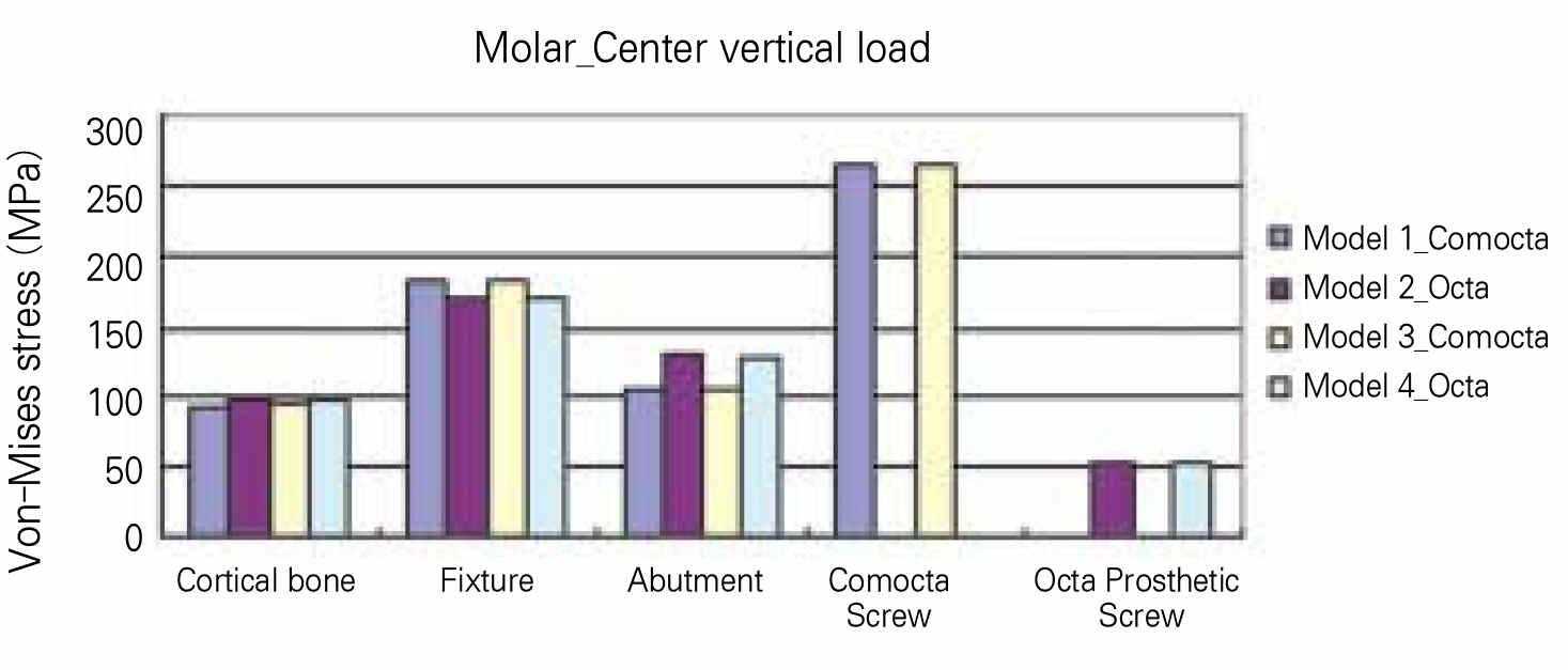
Fig. 4. Maximum stress of molar implant components under center vertical load.

Fig. 5. Maximum stress of premolar implant components under center vertical load.
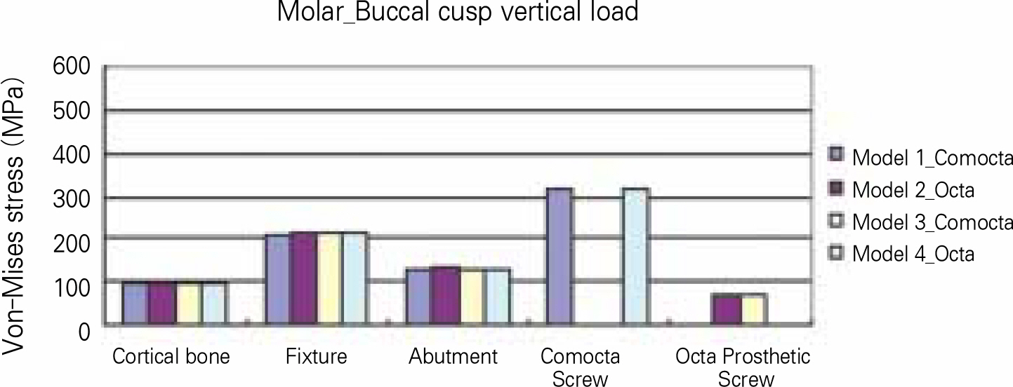
Fig. 6. Maximum stress of molar implant components under buccal cusp vertical load.
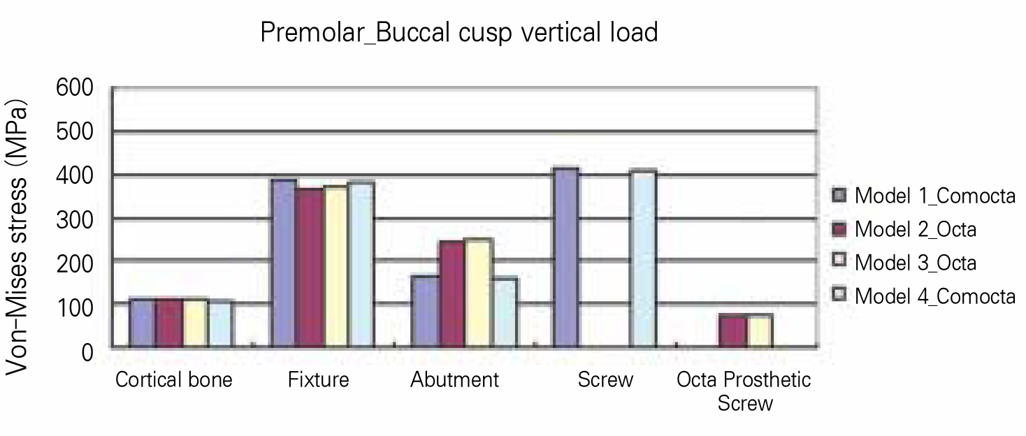
Fig. 7. Maximum stress of premolar implant components under buccal cusp vertical load.
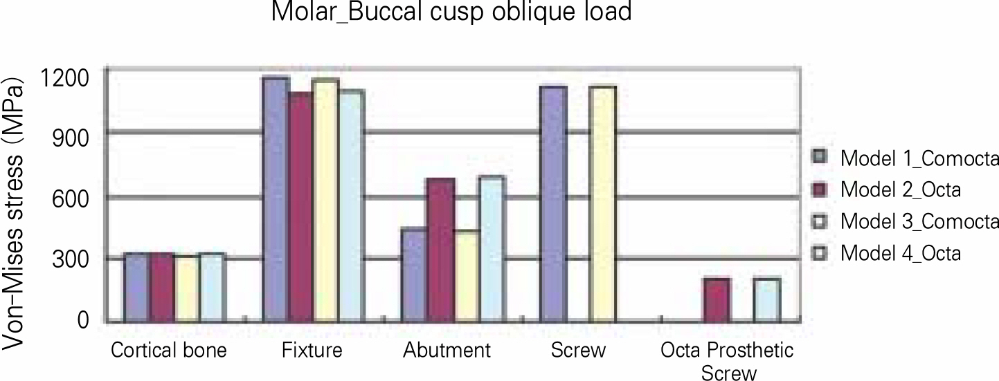
Fig. 8. Maximum stress of molar implant components under buccal cusp oblique load.
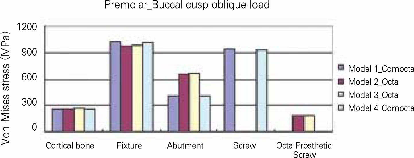
Fig. 9. Maximum stress of premolar implant components under buccal cusp oblique load.

Fig. 10. Stress distribution of 4 models under buccal cusp oblique load.

Fig. 11. Sectional maximum principle stress distribution under buccal cusp oblique load.
Reference
-
1.Adell R., Eriksson B., Lekholm U., Bra � nemark PI., Jemt T. Long-term follow-up study of osseointegrated implants in the treatment of totally edentulous jaws. Int J Oral Maxillofac Implants. 1990. 5:347–59.2.Albrektsson T. A multicenter report on osseointegrated oral implants. J Prosthet Dent. 1988. 60:75–84.
Article3.Preiskel HW., Tsolka P. The DIA anatomic abutment system and telescopic prostheses: a clinical report. Int J Oral Maxillofac Implants. 1997. 12:628–33.4.Singer A., Serfaty V. Cement-retained implant-supported fixed partial dentures: a 6-month to 3-year follow-up. Int J Oral Maxillofac Implants. 1996. 11:645–9.5.Misch CE. Screw-retained versus cement-retained implant-supported prostheses. Pract Periodontics Aesthet Dent. 1995. 7:15–8.6.Clelland NL., Carr AB., Gilat A. Comparison of strains transferred to a bone simulant between as-cast and postsol-dered implant frameworks for a five-implant-supported fixed prosthesis. J Prosthodont. 1996. 5:193–200.
Article7.English CE. Implant-supported versus implant-natural tooth-supported fixed partial dentures. J Dent Symp. 1993. 1:10–5.8.Khraisat A., Stegaroiu R., Nomura S., Miyakawa O. Fatigue resistance of two implant/abutment joint designs. J Prosthet Dent. 2002. 88:604–10.
Article9.Lee JM., Kim YS., Kim CW., Kim YH. 3-D FEA of three different single tooth abutments: Cement-retained vs screw-retained. J Korean Acad Prosthodont. 1999. 37:269–88.10.Guichet DL., Caputo AA., Choi H., Sorensen JA. Passivity of fit and marginal opening in screw- or cement-retained implant fixed partial denture designs. Int J Oral Maxillofac Implants. 2000. 15:239–46.11.Hebel KS., Gajjar RC. Cement-retained versus screw-retained implant restorations: Achieving optimal occlusion and esthetics in implant dentistry. J Prosthet Dent. 1997. 77:28–35.
Article12.Preiskel HW., Tsolka P. Telescopic prostheses for implants. Int J Oral Maxillofac Implants. 1998. 13:352–7.13.Lindstrom H., Preiskel H. The implant-supported telescopic prosthesis: a biomechanical analysis. Int J Oral Maxillofac Implants. 2001. 16:34–42.14.Preiskel H., Tsolka P. Cement-and screw-retained implant-supported prostheses: up to 10 years of follow-up of a new design. Int J Oral Maxillofac Implants. 2004. 19:87–91.15.Robert G. Restorative dental materials,. 9th ed. Mosby;1993. p. 54.16.Phillips RW. Skinner' s science of dental materials,. 8th ed. WB Saunders;1982. p. 55.17.Kenneth AJ. Phillips' science of dental materials,. 10th ed. WB Saunders;1996. p. 66.18.Cook SD., Weinstein AM., Klawitter JJ. A three-dimensional finite element analysis of a porous rooted Co-Cr-Mo alloy dental implant. J Dent Res. 1982. 61:25–9.19.Katona TR., Winkler MM. Stress analysis of a bulk-filled Class V light-cured composite restoration. J Dent Res. 1994. 73:1470–7.
Article20.Ko HJ., Chung CH. Finite element analysis of stresses induced by osseointegrated prostheses with or without connection between natural tooth and osseointegrated abutments. J Korean Acad Prosthodont. 1991. 29:147–60.21.Kim DW., Kim YS. A study on the osseointegrated prosthesis using three dimensional finite element method. J Korean Acad Prosthodont. 1991. 29:167–214.22.Squier RS., Psoter WJ., Taylor TD. Removal torques of conical, tapered implant abutments: the effects of anodization and reduction of surface area. Int J Oral Maxillofac Implants. 2002. 17:24–7.23.Lum LB., Osier JF. Load transfer from endosteal implants to supporting bone: an analysis using statics. Part one: Horizontal loading. J Oral Implantol. 1992. 18:343–8.24.Lum LB., Osier JF. Load transfer from endosteal implants to supporting bone: an analysis using statics. Part two: Axial loading. J Oral Implantol. 1992. 18:349–53.25.Clelland NL., Lee JK., Bimbenet OC., Gilat A. Use of an axisymmetric finite element method to compare maxillary bone variables for a loaded implant. J Prosthodont. 1993. 2:183–9.
Article26.Mo ¨llersten L., Lockowandt P., Linde ´ n LA. Comparison of strength and failure mode of seven implant systems: an in vitro test. J Prosthet Dent. 1997. 78:582–91.27.Norton MR. Assessment of cold welding properties of the internal conical interface of two commercially available implant systems. J Prosthet Dent. 1999. 81:159–66.
Article28.Boggan RS., Strong JT., Misch CE., Bidez MW. Influence of hex geometry and prosthetic table width on static and fatigue strength of dental implants. J Prosthet Dent. 1999. 82:436–40.
Article29.Holmes DC., Grigsby WR., Goel VK., Keller JC. Comparison of stress transmission in the IMZ implant system with polyoxymethylene or titanium intramobile element: a finite element stress analysis. Int J Oral Maxillofac Implants. 1992. 7:450–8.30.Kwon JH., Choi MH., Kim YL., Cho HW. Three-dimensional finite element stress analysis of single implant restoration using different fixture and abutment screw diameters. J Korean Acad Prosthodont. 2005. 43:105–19.
- Full Text Links
-
- Actions
-
Cited
- CITED
-
- Close
- Share
-
- Similar articles
-
- A Comparison Of Load Transfer In Screw-And Cement-Retained Implant Fixed Partial Denture Designs
- Finite element analysis of stresses and deflections induced by fixed partial denture using endosteal implant
- THREE-DIMENSIONAL FINI6E ELEMENT ANALYSIS OF THE ENDOSSEOUS IMPLANT DESIGNS
- The three dimensional finite element analysis of stress distribution and deformation in mandible according to the position of pontic in two implants supported three-unit fixed partial denture
- The three dimensional finite element analysis of the stress distribution according to the implant and thread designs
