J Korean Acad Prosthodont.
2010 Jul;48(3):194-201. 10.4047/jkap.2010.48.3.194.
Fracture resistance and marginal fidelity of zirconia crown according to the coping design and the cement type
- Affiliations
-
- 1Department of Prosthodontics, College of Dentistry, Wonkwang University, Gunpo, Korea. scoh@wku.ac.kr
- 2Implant Prosthodontics, Graduate School of Clinical Dentistry, Hallym University, Seoul, Korea.
- KMID: 2195555
- DOI: http://doi.org/10.4047/jkap.2010.48.3.194
Abstract
- PURPOSE
The purpose was to compare the marginal fidelity and the fracture resistance of the zirconia crowns according to the various coping designs with different thicknesses and cement types. Material and
METHODS
Zirconia copings were designed and fabricated with various thicknesses using the CAD/CAM system (Everest, KaVo Dental GmbH, Biberach., Germany). Eighty zirconia copings were divided into 4 groups (Group I: even 0.3 mm thickness, Group II: 0.3 mm thickness on the buccal surface and the buccal half of occlusal surface and the 0.6 mm thickness on the lingual surface and the lingual half of occlusal surface, Group III: even 0.6 mm thickness, Group IV: 0.6 mm thickness on the buccal surface and the buccal half of occlusal surface and the 1.0 mm thickness on the lingual surface and the lingual half of occlusal surface) of 20. By using a putty index, zirconia crowns with the same size and contour were fabricated. Each group was divided into two subgroups by type of cement: Cavitec(R) (Kerr Co, USA) and Panavia-F(R) (Kuraray Medical Inc, Japan). After the cementation of the crowns with a static load compressor, the marginal fidelity of the zirconia crowns were measured at margins on the buccal, lingual, mesial and distal surfaces, using a microscope of microhardness tester (Matsuzawa, MXT-70, Japan, x100). The fracture resistance of each crown was measured using a universal testing machine (Z020, Zwick, Germany) at a crosshead speed of 1 mm/min. The results were analyzed statistically by the two-way ANOVA and oneway ANOVA and Duncan's multiple range test at alpha= .05.
RESULTS
Group I and III showed the smallest marginal fidelity, while group II demonstrated the largest value in Cavitec(R) subgroup (P < .05). For fracture resistance, group III and IV were significantly higher than group I and II in Cavitec(R) subgroup (P < .05). The fracture resistances of Panavia-F(R) subgroup were not significantly different among the groups (P > .05). Panavia-F(R) subgroup showed significantly higher fracture resistance than Cavitec(R) subgroup in group I and II (P < .05).
CONCLUSION
Within the limitation of this study, considering fracture resistance or marginal fidelity and esthetics, a functional ceramic substructure design of the coping with slim visible surface can be used for esthetic purposes, or a thick invisible surface to support the veneering ceramic can be used depending on the priority.
Figure
-
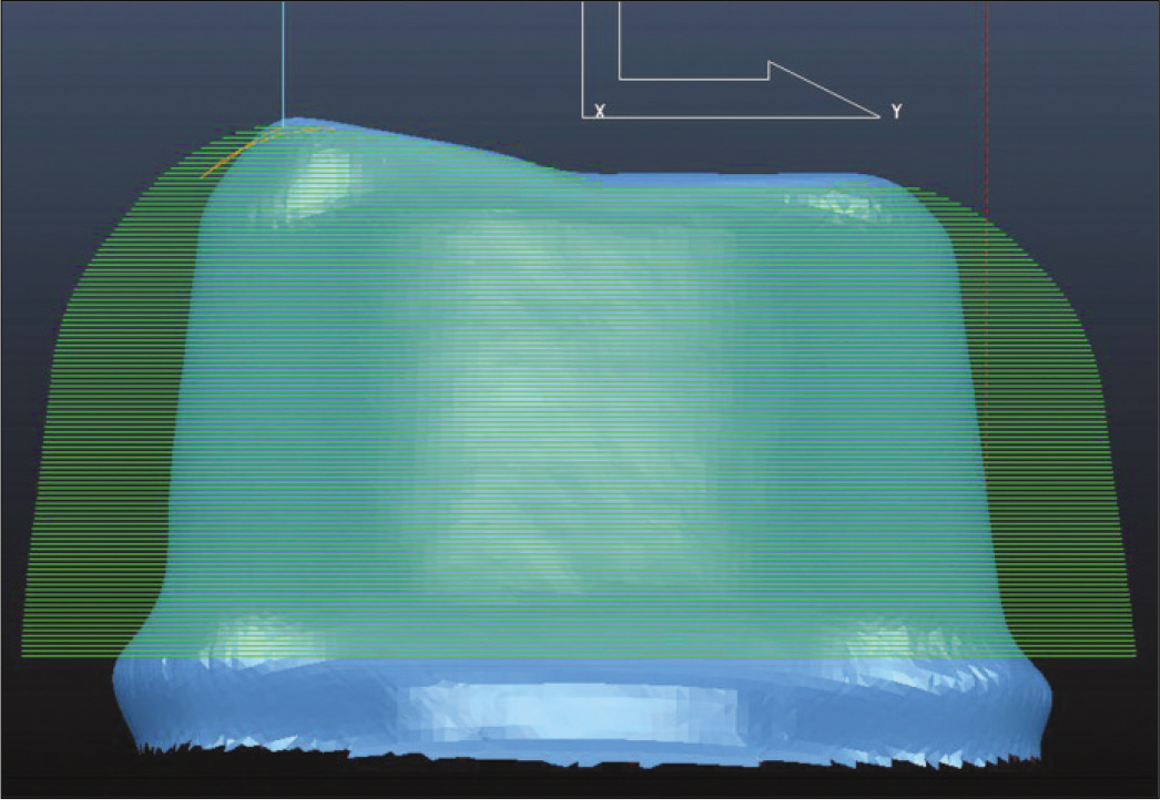
Fig. 1. 3D scanned image of stone model using the ISCAN D100® after modification.
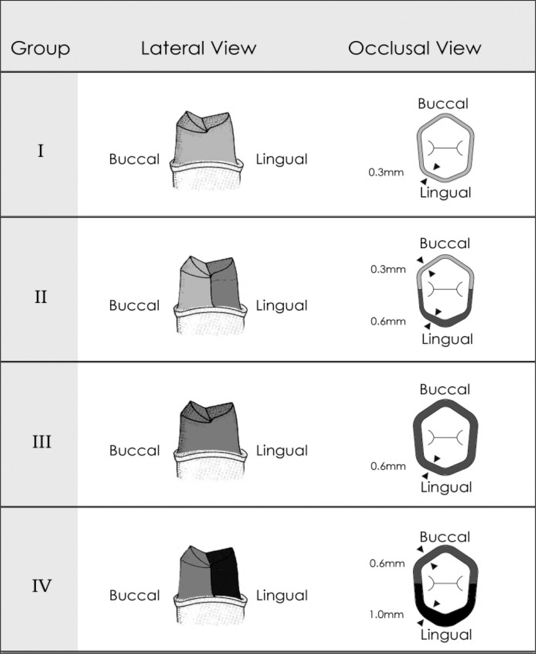
Fig. 2. Schematic diagram of lateral and occlusal view of zirconia coping according to the group.

Fig. 3. Assembly designed for measuring the fracture resistance of fabricated zirconia crown on a universal testing machine (A), and a schematic diagram of circled area (B).
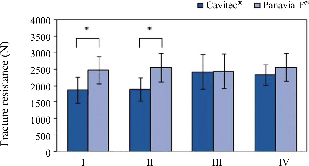
Fig. 4. Comparison of fracture resistances of zirconia crowns according to the cement type. ∗ means significant difference by t-test at α = .05.
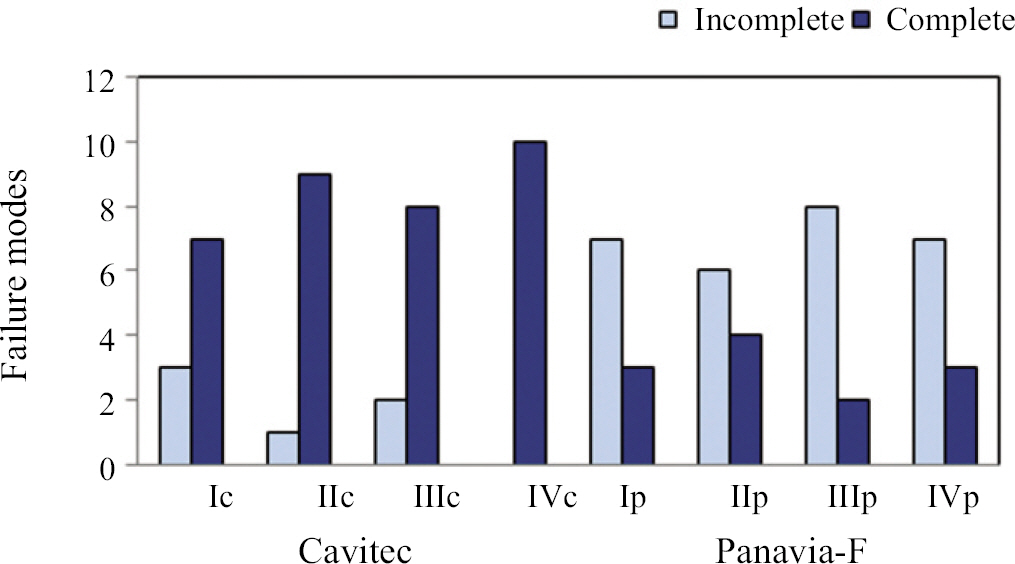
Fig. 5. Comparison of failure modes of zircornia crowns with different coping design and cement type. Ic, IIc, IIIc, IVc means group I, II, III, IV in Cavitec® Subgroup, Ip, IIp, IIIp, IVp means group I, II, III, IV in Panavia-F® subgroup.
Reference
-
1.Filser F., Kocher P., Weibel F., Lu ¨thy H., Scha ¨rer P., Gauckler LJ. Reliability and strength of all-ceramic dental restorations fabricated by direct ceramic machining (DCM). Int J Comput Dent. 2001. 4:89–106.2.Piconi C., Maccauro G. Zirconia as a ceramic biomaterial. Biomaterials. 1999. 20:1–25.
Article3.Tinschert J., Natt G., Mautsch W., Spiekermann H., Anusavice KJ. Marginal fit of alumina-and zirconia-based fixed partial dentures produced by a CAD/CAM system. Oper Dent. 2001. 26:367–74.4.Jeong HC. Fracture strength of zirconia monolithic crowns. J Korean Acad Prosthodont. 2006. 44:157–64.5.Kelly JR., Tesk JA., Sorensen JA. Failure of all-ceramic fixed partial dentures in vitro and in vivo: analysis and modeling. J Dent Res. 1995. 74:1253–8.
Article6.Fischer H., Weber M., Marx R. Lifetime prediction of all-ceramic bridges by computational methods. J Dent Res. 2003. 82:238–42.
Article7.Kupiec KA., Wuertz KM., Barkmeier WW., Wilwerding TM. Evaluation of porcelain surface treatments and agents for composite-to-porcelain repair. J Prosthet Dent. 1996. 76:119–24.
Article8.Ozcan M., Nijhuis H., Valandro LF. Effect of various surface conditioning methods on the adhesion of dual-cure resin cement with MDP functional monomer to zirconia after thermal aging. Dent Mater J. 2008. 27:99–104.9.Baek KH., Woo YH., Kwon KR., Kim HS. Spectrophotometric analysis of the influence to shade of zirconia core on the color of ceramic. J Korean Acad Prosthodont. 2008. 46:409–19.10.Shin MR., Kim MJ., Oh SC. Fracture load and marginal fitness of zirconia ceramic coping by design and coloration. J Korean Acad Prothodont. 2009. 47:406–15.
Article11.Besimo C., Jeger C., Guggenheim R. Marginal adaptation of titanium frameworks produced by CAD/CAM techniques. Int J Prosthodont. 1997. 10:541–6.12.May KB., Russell MM., Razzoog ME., Lang BR. Precision of fit: the Procera AllCeram crown. J Prosthet Dent. 1998. 80:394–404.
Article13.Kim DK., Cho IH., Lim JH., Lim HS. On the marginal fidelity of all-ceramic core using CAD/CAM system. J Korean Acad Prosthodont. 2003. 41:20–34.14.Nakamura T., Dei N., Kojima T., Wakabayashi K. Marginal and internal fit of Cerec 3 CAD/CAM all-ceramic crowns. Int J Prosthodont. 2003. 16:244–8.15.Grossman DG., Nelson JW. The bonded Dicor crown. IADR abstract no 800, J Dent Res. 1987. 66:206.16.Hobo S., Shillingburg HT Jr. Porcelain-fused to metal: tooth preparation and coping design. J Prosthet Dent. 1973. 30:28–36.17.Miller LL. Framework design in ceramo-metal restorations. Dent Clin North Am. 1977. 21:699–716.18.Groten M., Pro ¨bster L. The influence of different cementation modes on the fracture resistance of feldspathic ceramic crowns. Int J Prosthodont. 1997. 10:169–77.19.Eden GT., Kacicz JM. Dicor crown strength improvement due to bonding. IADR abstract no 801, J Dent Res. 1987. 66:207.20.Jung WY., Oh SC., Dong JK. A study on the microleakage of the ips empress ceramic crown according to margin types and resin cement. J Korean Acad Prosthodont. 1998. 36:789–804.21.Kern M., Thompson VP. Bonding to glass infiltrated alumina ceramic: adhesive methods and their durability. J Prosthet Dent. 1995. 73:240–9.
Article22.Yoon JT., Lee SH., Yang JH. The influence of surface treatments on the shear bond strength of resin cements to In-ceram core. J Korean Acad Prosthodont. 2000. 38:129–46.23.Lim JH. Marginal fidelity and fracture strength of In-ceram crowns according to various resin cements. J Korean Acad Prosthodont. 1998. 36:888–99.24.Strub JR., Beschnidt SM. Fracture strength of 5 different all-ceramic crown systems. Int J Prosthodont. 1998. 11:602–9.25.Castellani D., Baccetti T., Giovannoni A., Bernardini UD. Resistance to fracture of metal ceramic and all-ceramic crowns. Int J Prosthodont. 1994. 7:149–54.
- Full Text Links
-
- Actions
-
Cited
- CITED
-
- Close
- Share
-
- Similar articles
-
- MARGINAL FIDELITY AND FRACTURE STRENGTH OF IN-CERAM CROWNS ACCORDING TO VARIOUS RESIN CEMENTS
- A STUDY ON MARGINAL FIDELITY OF CERAMIC METAL COPINGS TREATED BY VARIOUS METHODS
- Biomechanical three-dimensional finite element analysis of monolithic zirconia crown with different cement type
- The marginal fidelity of Procera(R) AllCeram alumina copings and crowns of patients
- Fracture strength of zirconia monolithic crowns
