Considerations for increasing denture stability: a case report
- Affiliations
-
- 1Department of Prosthodontics, School of Dentistry, Kyung Hee University, Seoul, Korea. ahranp@khu.ac.kr
- KMID: 2195474
- DOI: http://doi.org/10.4047/jkap.2012.50.4.311
Abstract
- When wearing complete dentures, patients want to function naturally within a physiologically stable range. To do this, recovery of esthetics, biologically stable arrangement and contour, and occlusal contacts with denture stability are necessary. In this case report, a complete denture patient of adverse conditions was presented. To increase stability of the dentures, functional impression was made by border molding using the neutral zone. The dentures were checked for physiological centric relations and stable occlusion. The clinical results showed satisfactory results on function and esthetics.
Figure
-
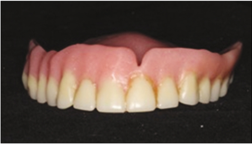
Fig. 1 Pre-existing old denture.
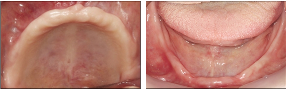
Fig. 2 Initial intra-oral view.
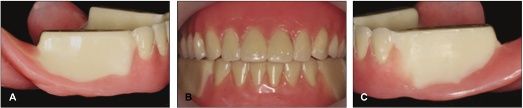
Fig. 3 Treatment denture. (A) the right side (B) the frontal side (C) the left side.
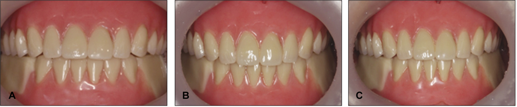
Fig. 4 After treatment denture delivery. (A) treatment denture delivery, (B) next day ; mid-line deviation and premature contact were appeared. So, occlusal adjustment was made. (C) next week; the dentures were checked for physiological centric relations.
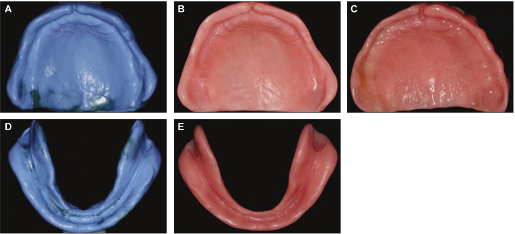
Fig. 5 Comparison of the inner surface after a final impression. (A) final impression surface (upper), (B) final denture inner surface(upper), (C) pre-existing old denture inner surface, (D) final impression surface(lower), (E) final denture inner surface (lower).
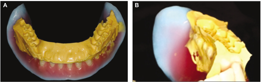
Fig. 6 Neutral zone checked and modified 0.5 mm to the outside. (A) neutral zone checked, (B) left tongue space invasion.
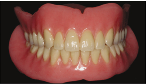
Fig. 7 The final denture delivery after occlusal adjustment.

Fig. 8 Three pairs of dentures replicated with different posterior occlusions. (A) normal occlusion, (B) lingualized occlusion, (C) 0° teeth occlusion.
Reference
-
1. Darvell BW, Clark RK. The physical mechanisms of complete denture retention. Br Dent J. 2000. 189:248–252.
Article2. Jacob RF. The traditional therapeutic paradigm: complete denture therapy. J Prosthet Dent. 1998. 79:6–13.
Article3. Ahmad AJ, Peter JN. Neutral zone in complete dentures; systematic analysis of evidence and technique. Smile Dent J. 2011. 6:8–12.4. McGarry TJ, Nimmo A, Skiba JF, Ahlstrom RH, Smith CR, Koumjian JH. Classification system for complete edentulism. Dent Today. 2001. 20:90–95.
Article5. Inada M, Yamazaki T, Shinozuka O, Sekiguchi G, Tamamori Y, Ohyama T. Complete denture treatments for a cerebral palsy patient by using a treatment denture. A case report. J Med Dent Sci. 2002. 49:171–177.6. Zuccolotto MC, Nóbilo KA, Nunes Lde J, Hotta TH. Sliding plates on complete dentures as a treatment of temporomandibular disorder: a case report. Cranio. 1999. 17:289–292.
Article7. Duncan JP, Raghavendra S, Taylor TD. A selective-pressure impression technique for the edentulous maxilla. J Prosthet Dent. 2004. 92:299–301.
Article8. Ladha K, Gill S, Gupta R, Verma M. Neutral zone approach for the rehabilitation of a severely atrophic mandibular ridge: Case report. Gen Dent. 2012. 60:e166–e169.9. Ortman HR. The role of occlusion in preservation and prevention in complete denture prosthodontics. J Prosthet Dent. 1971. 25:121–138.
Article10. Levin B. A review of artificial posterior tooth forms including a preliminary report on a new posterior tooth. J Prosthet Dent. 1977. 38:3–15.
Article11. Boretti G, Bickel M, Geering AH. A review of masticatory ability and efficiency. J Prosthet Dent. 1995. 74:400–403.
Article12. Sato S, Nasu F, Motegi K. Analysis of kinesiograph recordings and masticatory efficiency after treatment of non-reducing disk displacement of the temporomandibular joint. J Oral Rehabil. 2003. 30:708–713.
Article13. Sierpińska T, Gołebiewska M, Długosz JW. The relationship between masticatory efficiency and the state of dentition at patients with non rehabilitated partial lost of teeth. Adv Med Sci. 2006. 51:196–199.14. Escudeiro Santos C, de Freitas O, Spadaro AC, Mestriner-Junior W. Development of a colorimetric system for evaluation of the masticatory efficiency. Braz Dent J. 2006. 17:95–99.
Article15. Schimmel M, Christou P, Herrmann F, Müller F. A two-colour chewing gum test for masticatory efficiency: development of different assessment methods. J Oral Rehabil. 2007. 34:671–678.
Article16. Nokubi T, Nokubi F, Yoshimuta Y, Ikebe K, Ono T, Maeda Y. Measuring masticatory performance using a new device and β-carotene in test gummy jelly. J Oral Rehabil. 2010. 37:820–826.
Article17. Maria EOA, Wilson M Jr, Fábio RD, Cícero RGN, Samira AS. Clinical assessment of masticatory efficiency in the rehabilitation of edentulous patients. Braz J Oral Sci. 2011. 10:217–220.18. Kwon KR, Park NS, Choi DG. A study on masticatory performance and function by posterior occlusal schemes in complete denture. J Korean Acad Prosthodont. 1996. 34:539–573.19. Carlsson GE. Early in contrast to recent methods to evaluate masticatory function in implant patients. J Prosthodont Res. 2012. 56:3–10.
Article
- Full Text Links
-
- Actions
-
Cited
- CITED
-
- Close
- Share
-
- Similar articles
-
- Comparative analysis of case series for the prosthetic rehabilitation of edentulous patients using suction denture
- Effects and clinical considerations of denture adhesives
- Palate bone exposure from flexible denture: a case report
- Complete denture fabricated by Jiro Abe's method for edentulous patient with severe alveolar ridge resorption: a case report
- Clinical application of mandibular removable partial denture using implant-supported surveyed crown: A case report
