Update in surgical treatment of shoulder injuries
- Affiliations
-
- 1CM General Hospital, Seoul, Korea. sanghoon.lhee@gmail.com
- KMID: 2194917
- DOI: http://doi.org/10.5124/jkma.2014.57.8.667
Abstract
- Increased life expectancy, combined with advancements in medicine, has given rise to an increased prevalence of shoulder injuries. Additionally, the recent social trend in Korea of participation of younger generations in sports activities has also contributed to this increased prevalence. Many healthcare institutions, however, are performing surgery for shoulder injuries without thorough consideration of the complete clinical picture. Only a few of these injuries require surgery, and most of them can be treated and improved with conservative management. The need for surgery should be decided in conjunction with the consideration of the patient's age, living environment and level of physical activity. Here, we give a brief introduction to indications and methods for surgical treatment of shoulder injuries such as rotator cuff injuries, calcifying tendinitis, shoulder instability, osteoarthritis of the glenohumeral joint, and adhesive capsulitis.
Keyword
MeSH Terms
Figure
-

Figure 1 (A) Normal shoulder anatomy. (B) Adhesive capsulitis anatomy.

Figure 2 Arthroscopic capsular release. Thickened capsule.
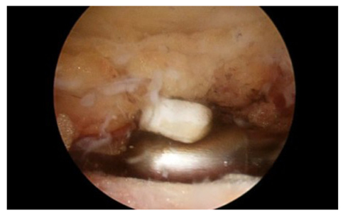
Figure 3 Subacromial bursitis.

Figure 4 Open acromioplasty.
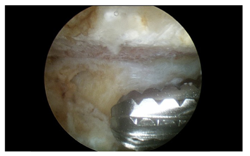
Figure 5 Arthroscopic acromioplasty.
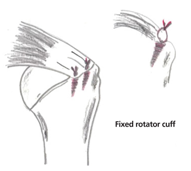
Figure 6 Rotator cuff repair using suture-anchor.
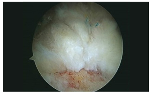
Figure 7 Single-row rotator cuff repair.
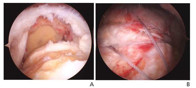
Figure 8 (A) Massive rotator cuff tear. (B) Suture-bridge repair of rotator cuff.
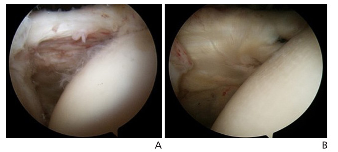
Figure 9 (A) Subscapularis tear. (B) Arthroscopic subscapularis repair based on Lhee's technique.
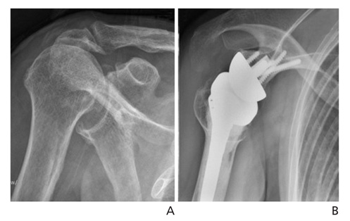
Figure 10 (A) Cuff tear arthropathy . (B) Reverse total shoulder arthroplasty state.
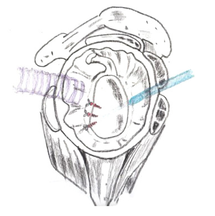
Figure 11 Schematic image of arthroscopic Bankart repair.
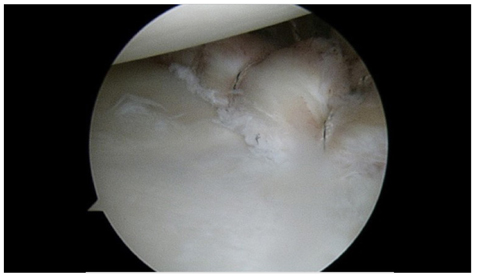
Figure 12 Arthroscopic Bankart repair state.
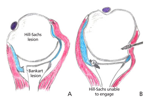
Figure 13 (A) Bankart lesion & Hill-Sachs lesion. (B) Bankart repair state and Remplissage.

Figure 14 Arthroscopic remplissage procedure.
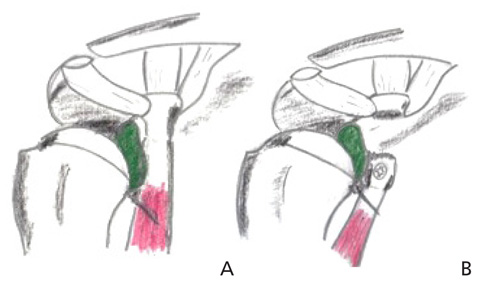
Figure 15 (A) Preoperative state of Latarjet procedure. (B) Postoperative state of Latarjet procedure.

Figure 16 Latarjet procudure postoperative X-ray. (A) Scapular-Y view, (B) anteroposterior view and (C) axial view.
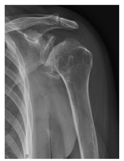
Figure 17 Anteroposterior view of glenohumeral joint arthritis.
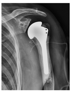
Figure 18 Total shoulder arthroplasty.
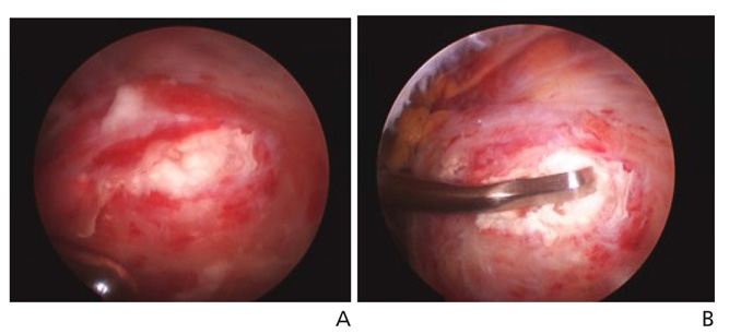
Figure 19 (A) Calcific deposit in rotator cuff tendon. (B) Decompression of calcium.
Reference
-
1. Neer CS 2nd. Anterior acromioplasty for the chronic impingement syndrome in the shoulder: a preliminary report. J Bone Joint Surg Am. 1972; 54:41–50.
Article2. Lhee SH, Kho DH, Park JY. Shoulder operation. J Korean Med Assoc. 2009; 52:795–804.
Article3. Griggs SM, Ahn A, Green A. Idiopathic adhesive capsulitis. A prospective functional outcome study of nonoperative treatment. J Bone Joint Surg Am. 2000; 82:1398–1407.4. Griesser MJ, Harris JD, Campbell JE, Jones GL. Adhesive capsulitis of the shoulder: a systematic review of the effectiveness of intra-articular corticosteroid injections. J Bone Joint Surg Am. 2011; 93:1727–1733.5. Hsu JE, Anakwenze OA, Warrender WJ, Abboud JA. Cur-rent review of adhesive capsulitis. J Shoulder Elbow Surg. 2011; 20:502–514.
Article6. Gobezie R, Pacheco IH, Petit CJ, Millett PJ. Dislocation and instability after arthroscopic capsular release for refractory frozen shoulder. Am J Orthop (Belle Mead NJ). 2007; 36:672–674.7. Papadonikolakis A, McKenna M, Warme W, Martin BI, Matsen FA 3rd. Published evidence relevant to the diagnosis of impingement syndrome of the shoulder. J Bone Joint Surg Am. 2011; 93:1827–1832.
Article8. Yamaguchi K, Tetro AM, Blam O, Evanoff BA, Teefey SA, Middleton WD. Natural history of asymptomatic rotator cuff tears: a longitudinal analysis of asymptomatic tears detected sonographically. J Shoulder Elbow Surg. 2001; 10:199–203.
Article9. Duquin TR, Buyea C, Bisson LJ. Which method of rotator cuff repair leads to the highest rate of structural healing? A systematic review. Am J Sports Med. 2010; 38:835–841.
Article10. Mascarenhas R, Chalmers PN, Sayegh ET, Bhandari M, Verma NN, Cole BJ, Romeo AA. Is double-row rotator cuff repair clinically superior to single-row rotator cuff repair: a systematic review of overlapping meta-analyses. Arthroscopy. 2014; 05. 09. [Epub]. http://dx.doi.org/10.1016/j.arthro.2014.03.015.11. Ricchetti ET, Aurora A, Iannotti JP, Derwin KA. Scaffold devices for rotator cuff repair. J Shoulder Elbow Surg. 2012; 21:251–265.
Article12. Derwin KA, Badylak SF, Steinmann SP, Iannotti JP. Extracellular matrix scaffold devices for rotator cuff repair. J Shoulder Elbow Surg. 2010; 19:467–476.
Article13. Lhee SH, Park JY. Prospective randomized comparative study of 191 subscapularis tear: clinical and radiologic outcome: arthroscopic repair vs. debridement. J Shoulder Elbow Surg. 2013; 22:e24–e26.14. Adams CR, Schoolfield JD, Burkhart SS. The results of arthroscopic subscapularis tendon repairs. Arthroscopy. 2008; 24:1381–1389.
Article15. Walker M, Brooks J, Willis M, Frankle M. How reverse shoulder arthroplasty works. Clin Orthop Relat Res. 2011; 469:2440–2451.
Article16. Ticker JB, Bigliani LU, Soslowsky LJ, Pawluk RJ, Flatow EL, Mow VC. Inferior glenohumeral ligament: geometric and strain-rate dependent properties. J Shoulder Elbow Surg. 1996; 5:269–279.
Article17. Young AA, Maia R, Berhouet J, Walch G. Open Latarjet procedure for management of bone loss in anterior instability of the glenohumeral joint. J Shoulder Elbow Surg. 2011; 20:2 Suppl. S61–S69.
Article18. Uhthoff HK, Loehr JW. Calcific tendinopathy of the rotator cuff: pathogenesis, diagnosis, and management. J Am Acad Orthop Surg. 1997; 5:183–191.
Article
- Full Text Links
-
- Actions
-
Cited
- CITED
-
- Close
- Share
-
- Similar articles
-
- Diagnosis for Acute Traumatic Shoulder Injuries
- Recent Updates Regarding Outcomes and Complications of Reverse Total Shoulder Arthroplasty
- Surgical treatment of shoulder pain: a focused study on rotator cuff injury
- Rehabilitation of Sports Related Shoulder Injuries
- The Treatment of Acromioclavicular Separation
