Hemangioma of the Flexor Digitorum Superficialis Tendon Sheath in the Hand
- Affiliations
-
- 1Department of Orthopaedic Surgery, Samsung Changwon Hospital, Sungkyunkwan University School of Medicine, Changwon, Korea. oskimck@naver.com
- KMID: 2194138
- DOI: http://doi.org/10.12790/jkssh.2014.19.3.154
Abstract
- Hemangioma is a common tumor which can occur in any part of the body. It can develop in any area the hand. Hemangioma of the hand usually presents with swelling, pressure pain, accompanied by motion restriction. Rarely it has throbbing pain. We report the case of a 21-year-old woman who had a hemangioma of flexor digitorum superficialis tendon sheath in second finger with restriction of motion and were treated by surgical intervention.
Keyword
Figure
-

Fig. 1. (A) Axial view T1-weighted image shows that the signal of the lesion is equivalent to that of adjacent soft tissues. And T2-weighted image shows slightly ill-defined, lobulating slightly high signal intensity mass at the volar aspect of the proximal to middle phalanges level. (B) Sagittal view T1-weighted image shows ill-defined mixed high signal intensity nodule at the proximal phalanx level. And T2-weighted image shows that the high signal mass exist between bone and flexor tendons at the proximal phalanx level.
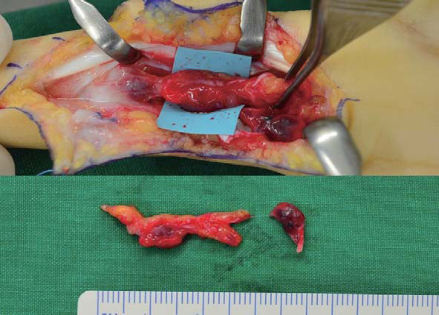
Fig. 2. Intraoperative findings: the 2.9×0.6 cm sized fresh red tumor is identified between second proximal phalanx bone and flexor digitorum superficialis, and it is attached to tendon sheath and vincula longa distally.

Fig. 3. Histological photograph (H&E). (A) The specimen shows that it is a cavernous type hemangioma (×40). (B) Vascular spaces filled with erythrocytes and fibrin clot (×100).
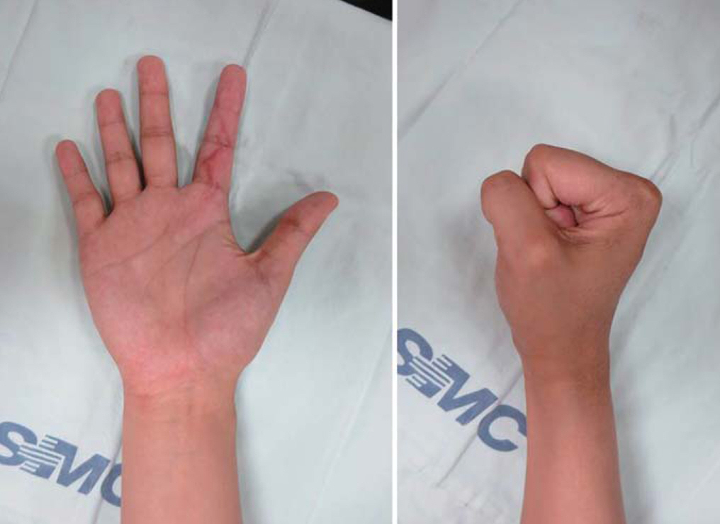
Fig. 4. Postoperative photographs show full flexion and full extension of left second finger.
Cited by 1 articles
-
Microscopic Decompression of Digital Nerve Surrounded by Hemangioma: A Case Report
Jun Gul Ko, Jun Hyeok Kim, Eun Young Rha, Jun Yong Lee, Gyeol Yoo, Sang Oon Baek
Arch Hand Microsurg. 2018;23(4):301-305. doi: 10.12790/ahm.2018.23.4.301.
Reference
-
1. Devaney K, Vinh TN, Sweet DE. Synovial hemangioma: a report of 20 cases with differential diagnostic considerations. Hum Pathol. 1993; 24:737–45.
Article2. Chung DW, Lee JH, Lee JH. Intramuscular hemangioma of lumbrical muscle: a case report. J Korean Soc Surg Hand. 2002; 7:203–7.3. Heo YM. Hemangioma of the flexor digitorum profundus of the hand: a case report. J Korean Soc Surg Hand. 2008; 13:142–5.4. Lalonde D, Martin A. Epinephrine in local anesthesia in finger and hand surgery: the case for wide-awake anesthesia. J Am Acad Orthop Surg. 2013; 21:443–7.
Article5. Stack HG. Tumours of the Hand. Postgrad Med J. 1964; 40:290–8.
Article6. George R, Lee K. Synovial angioma of the FDP flexor sheath: a rare cause of carpal tunnel syndrome. Open Orthop J. 2013; 7:72–4.
Article7. Waddell GF. A hemangioma involving tendons. J Bone Joint Surg Br. 1967; 49:138–41.8. Talwalkar S, Hayton M, Stilwell J, Temperley D, Freemont A. Tenosynovial haemangioma of the finger. Acta Orthop Belg. 2005; 71:618–21.9. Rico AA, Holguin PH, Gonzalez IG, Coba JM. Flexor tendon synovial sheath haemangioma mimicking subacute tenosynovitis. J Hand Surg Br. 1994; 19:704–5.
Article10. Spinner M, Moon S, Young L. Recurrent cavernous haemangioma of the extensor tendons of the hand. Hand. 1983; 15:223–7.
Article
- Full Text Links
-
- Actions
-
Cited
- CITED
-
- Close
- Share
-
- Similar articles
-
- A Case of Trigger Finger Following Longitudinal Tear of Flexor Digitorum Superficialis after Repeated Closed Injury
- Carpal Tunnel Syndrome Caused by Anatomic Variation of Flexor Digitorum Superficialis of Little Finger
- Primary Ring Flexor Digitorum Superficialis Transfer with Open Carpal Tunnel Release in Extreme Carpal Tunnel Syndrome
- Removal of a Movable Foreign Body in the Flexor Tendon Sheath of the Hand: A Case Report
- Concomitant Variations in Flexor Digitorum Superficialis: A Case Report
