Injury of the Arcuate Fasciculus in the Nondominant Hemisphere by Subfalcine Herniation in Patients with Intracerebral Hemorrhage : Two Case Reports and Literature Review
- Affiliations
-
- 1Department of Physical Medicine and Rehabilitation, College of Medicine, Yeungnam University, Daegu, Korea.
- 2Department of Neurosurgery, College of Medicine, Yeungnam University, Daegu, Korea.
- 3Department of Physical Medicine and Rehabilitation, Asan Medical Center, University of Ulsan College of Medicine, Seoul, Korea. wheel633@hanmail.net
- KMID: 2192112
- DOI: http://doi.org/10.3340/jkns.2016.59.3.306
Abstract
- Using diffusion tensor tractography (DTT), we demonstrated injury of the arcuate fasciculus (AF) in the nondominant hemisphere in two patients who showed subfalcine herniation after intracerebral hemorrhage (ICH) in the dominant hemisphere. Two patients (patient 1 and patient 2) with ICH and six age-matched control patients who have ICH on the left corona radiata and basal ganglia without subfalcine herniation were recruited for this study. DTT was performed at one month after onset in patient 1 and patient 2. AFs of both hemispheres in both patients were disrupted between Wernicke's and Broca's areas. The fractional anisotropy value and tract numbers of the right AFs in both patients were found to be more than two standard deviations lower than those of control patients. In contrast, the apparent diffusion coefficient value was more than two standard deviations higher than those of control patients. Using the configuration and parameters of DTT, we confirmed injury of the AF in the nondominant hemisphere in two patients with subfalcine herniation following ICH in the dominant hemisphere. Therefore, DTT would be a useful tool for detection of underlying injury of the AF in the nondominant hemisphere in patients with subfalcine herniation.
Keyword
Figure
-
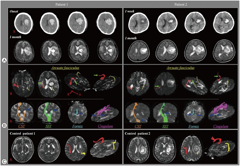
Fig. 1 A : Brain computed tomography images show an intracranial hemorrhage in the left corona radiata and basal ganglia and a left-to-right midline shift. T-2 weighted brain images show a leukomalatic lesion in the left corona radiata and basal ganglia. B : Diffusion tensor tractography (DTT) for the patient 1 and patient 2 shows disruptions of the arcuate fasciculus (AF) in both hemispheres. The right arcuate fasciculus in patient 1 and patient 2 are disrupted around the middle and posterior portions of the horizontal portion, respectively (arrow). The left arcuate fasciculus in both patients is disrupted at the junction between the ascending and horizontal portions. C : DTT for the control patients showed the preservation of the right AFs and the discontinuation of the left AFs after originating from Wernicke area.
Reference
-
1. Adams RD, Victor M, Ropper AH. Principles of neurology. ed 6. New York: McGraw-Hill;1997.2. Assaf Y, Pasternak O. Diffusion tensor imaging (DTI)-based white matter mapping in brain research : a review. J Mol Neurosci. 2008; 34:51–61. PMID: 18157658.
Article3. Bartha L, Benke T. Acute conduction aphasia : an analysis of 20 cases. Brain Lang. 2003; 85:93–108. PMID: 12681350.4. Byard RW. Patterns of cerebral and cerebellar herniation. Forensic Sci Med Pathol. 2013; 9:260–264. PMID: 22544455.
Article5. Catani M, Mesulam M. The arcuate fasciculus and the disconnection theme in language and aphasia : history and current state. Cortex. 2008; 44:953–961. PMID: 18614162.
Article6. Duffau H, Capelle L, Sichez N, Denvil D, Lopes M, Sichez JP, et al. Intraoperative mapping of the subcortical language pathways using direct stimulations. An anatomo-functional study. Brain. 2002; 125(Pt 1):199–214. PMID: 11834604.
Article7. Heiss WD, Kessler J, Thiel A, Ghaemi M, Karbe H. Differential capacity of left and right hemispheric areas for compensation of poststroke aphasia. Ann Neurol. 1999; 45:430–438. PMID: 10211466.
Article8. Heiss WD, Thiel A. A proposed regional hierarchy in recovery of post-stroke aphasia. Brain Lang. 2006; 98:118–123. PMID: 16564566.
Article9. Hong JH, Kim SH, Kim OL, Byun WM, Jang SH. Neural tract injuries by brain herniations after head trauma. J Head Trauma Rehabil. 2012; 27:154–158. PMID: 21386711.
Article10. Hosomi A, Nagakane Y, Yamada K, Kuriyama N, Mizuno T, Nishimura T, et al. Assessment of arcuate fasciculus with diffusion-tensor tractography may predict the prognosis of aphasia in patients with left middle cerebral artery infarcts. Neuroradiology. 2009; 51:549–555. PMID: 19434402.
Article11. Kim H, Na DL. Normative data on the Korean version of the Western Aphasia Battery. J Clin Exp Neuropsychol. 2004; 26:1011–1020. PMID: 15590457.
Article12. Kim SH, Jang SH. Prediction of aphasia outcome using diffusion tensor tractography for arcuate fasciculus in stroke. AJNR Am J Neuroradiol. 2013; 34:785–790. PMID: 23042924.
Article13. Lee AY, Shin DG, Park JS, Hong GR, Chang PH, Seo JP, et al. Neural tracts injuries in patients with hypoxic ischemic brain injury : diffusion tensor imaging study. Neurosci Lett. 2012; 528:16–21. PMID: 22982143.
Article14. Neil JJ. Diffusion imaging concepts for clinicians. J Magn Reson Imaging. 2008; 27:1–7. PMID: 18050325.
Article15. Nucifora PG, Verma R, Melhem ER, Gur RE, Gur RC. Leftward asymmetry in relative fiber density of the arcuate fasciculus. Neuroreport. 2005; 16:791–794. PMID: 15891571.
Article16. Payabvash S, Kamalian S, Fung S, Wang Y, Passanese J, Kamalian S, et al. Predicting language improvement in acute stroke patients presenting with aphasia : a multivariate logistic model using location-weighted atlas-based analysis of admission CT perfusion scans. AJNR Am J Neuroradiol. 2010; 31:1661–1668. PMID: 20488905.
Article17. Rosner MJ, Rosner SD, Johnson AH. Cerebral perfusion pressure : management protocol and clinical results. J Neurosurg. 1995; 83:949–962. PMID: 7490638.
- Full Text Links
-
- Actions
-
Cited
- CITED
-
- Close
- Share
-
- Similar articles
-
- Brain Lesions in Conduction Aphasia
- Spontaneous Spinal Subdural and Subarachnoid Hemorrhage with Concomitant Intracerebral Hemorrhage: A Case Report
- Repeated Intracerebral Hemorrhage from Developmental Venous Anomaly Alone
- Simultaneous Occurrence of Aneurysmal Subarachnoid Hemorrhage and Hypertensive Intracerebral Hemorrhage
- Injury of the Lower Ascending Reticular Activating System by Subfalcine Herniation in a Patient With a Cerebral Infarct
