A Morphometric Study of the Obturator Nerve around the Obturator Foramen
- Affiliations
-
- 1Department of Neurosurgery, Soonchunhyang University Gumi Hospital, Gumi, Korea. s90846@schmc.ac.kr
- KMID: 2192107
- DOI: http://doi.org/10.3340/jkns.2016.59.3.282
Abstract
OBJECTIVE
Obturator neuropathy is a rare condition. Many neurosurgeons are unfamiliar with the obturator nerve anatomy. The purpose of this study was to define obturator nerve landmarks around the obturator foramen.
METHODS
Fourteen cadavers were studied bilaterally to measure the distances from the nerve root to relevant anatomical landmarks near the obturator nerve, including the anterior superior iliac spine (ASIS), the pubic tubercle, the inguinal ligament, the femoral artery, and the adductor longus.
RESULTS
The obturator nerve exits the obturator foramen and travels infero-medially between the adductors longus and brevis. The median distances from the obturator nerve exit zone (ONEZ) to the ASIS and pubic tubercle were 114 mm and 30 mm, respectively. The median horizontal and vertical distances between the pubic tubercle and the ONEZ were 17 mm and 27 mm, respectively. The shortest median distance from the ONEZ to the inguinal ligament was 19 mm. The median inguinal ligament lengths from the ASIS and the median pubic tubercle to the shortest point were 103 mm and 24 mm, respectively. The median obturator nerve lengths between the ONEZ and the adductor longus and femoral artery were 41 mm and 28 mm, respectively.
CONCLUSION
The obturator nerve exits the foramen 17 mm and 27 mm on the horizontal and sagittal planes, respectively, from the pubic tubercle below the pectineus muscle. The shallowest area is approximately one-fifth medially from the inguinal ligament. This study will help improve the accuracy of obturator nerve surgeries to better establish therapeutic plans and decrease complications.
Figure
-
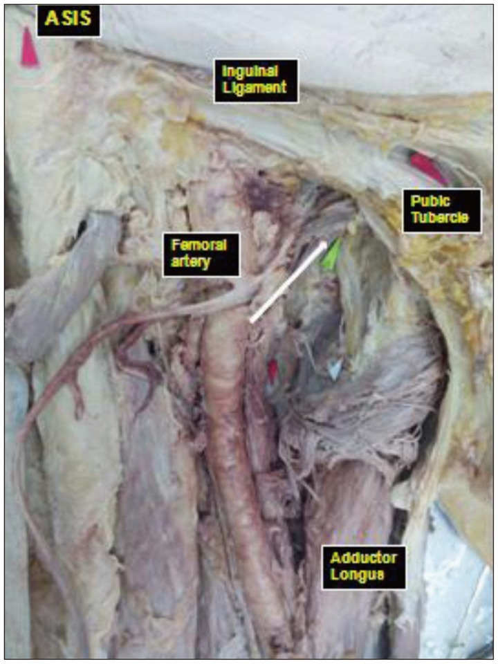
Fig. 1 Photograph of a cadaver specimen showing an anteroposterior view of the right thigh soft tissue dissection. The obturator nerve exit zone of the foramen (white arrow) was exposed after the pectineus muscle was dissected.
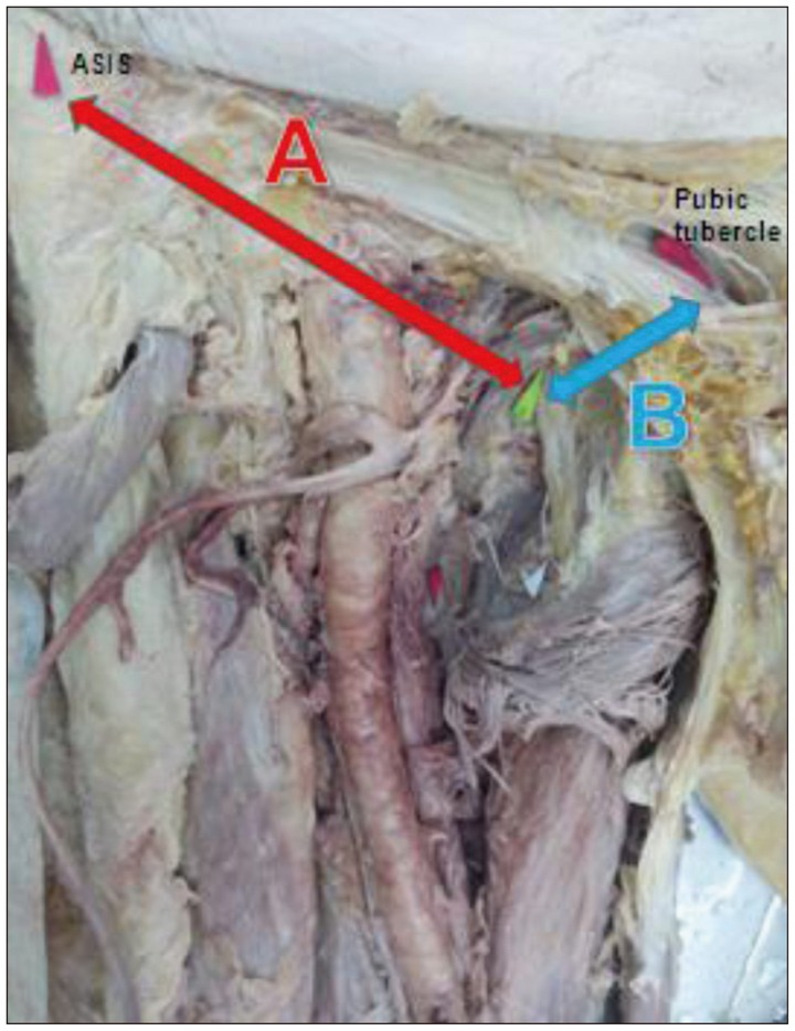
Fig. 2 Photograph of a cadaver specimen showing an anteroposterior view of the right thigh between the ASIS, the pubic tubercle, and the obturator nerve exit zone of the foramen after soft tissue dissection. A : the distance from the ASIS to the obturator nerve exit zone of the foramen, B : the distance from the pubic tubercle to the obturator nerve exit zone of the foramen. ASIS : anterior superior iliac spine
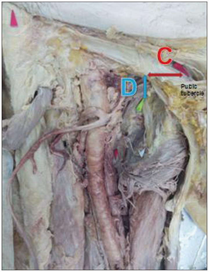
Fig. 3 Photograph of a cadaver specimen showing an anteroposterior view of the right thigh between the pubic tubercle and the obturator nerve exit zone of the foramen after soft tissue dissection. C : the horizontal distance, D : the vertical distance.

Fig. 4 Photograph of a cadaver specimen showing an anteroposterior view of the right thigh between the inguinal ligament and the obturator nerve exit zone of the foramen after soft tissue dissection. E : the shortest distance from the obturator nerve exit zone of the foramen to the inguinal ligament, F : the inguinal ligament length from the ASIS to point E, G : the inguinal ligament length from the pubic tubercle to point E. ASIS : anterior superior iliac spine.
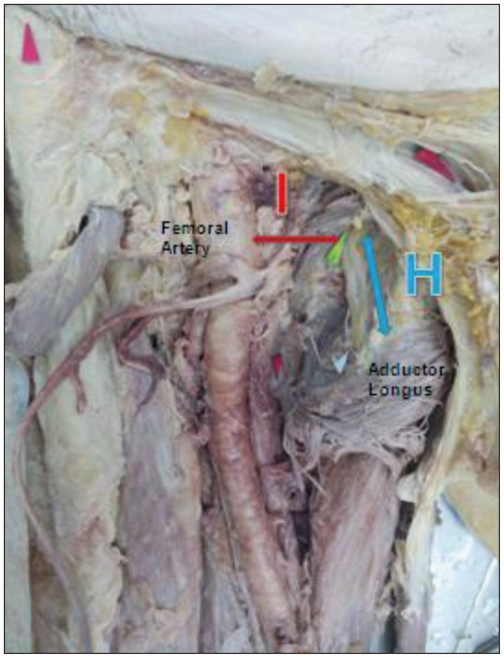
Fig. 5 Photograph of a cadaver specimen showing an anteroposterior view of the right thigh between the femoral artery and the obturator nerve exit zone of the foramen after soft tissue dissection. H : the obturator nerve length was exposed between the obturator nerve exit zone of the foramen and the adductor longus, I : the shortest distance from the obturator nerve exit zone of the foramen to the femoral artery.
Reference
-
1. Ahmadian A, Abel N, Uribe JS. Functional recovery of severe obturator and femoral nerve injuries after lateral retroperitoneal transpsoas surgery. J Neurosurg Spine. 2013; 18:409–414. PMID: 23432325.
Article2. Anagnostopoulou S, Kostopanagiotou G, Paraskeuopoulos T, Chantzi C, Lolis E, Saranteas T. Anatomic variations of the obturator nerve in the inguinal region : implications in conventional and ultrasound regional anesthesia techniques. Reg Anesth Pain Med. 2009; 34:33–39. PMID: 19258986.3. Choi EJ, Byun JM, Nahm FS, Lee PB. Obturator nerve block with botulinum toxin type B for patient with adductor thigh muscle spasm -a case report-. Korean J Pain. 2011; 24:164–168. PMID: 21935496.
Article4. Freisburger C, Nachtigall B, Wulf H. [Obturator nerve block]. Anasthesiol Intensivmed Notfallmed Schmerzther. 2010; 45:314–315. PMID: 20455186.5. Ghai A, Sangwan SS, Hooda S, Kiran S, Garg N. Obturator neurolysis using 65% alcohol for adductor muscle spasticity. Saudi J Anaesth. 2012; 6:282–284. PMID: 23162405.
Article6. Hadley G. Essential clinical anatomy. J Anat. 2007; 211:413.
Article7. Joniau SG, Van Baelen AA, Hsu CY, Van Poppel HP. Complications and functional results of surgery for locally advanced prostate cancer. Adv Urol. 2012; 2012:706309. PMID: 22291698.
Article8. Kim SH, Seok H, Lee SY, Park SW. Acetabular paralabral cyst as a rare cause of obturator neuropathy : a case report. Ann Rehabil Med. 2014; 38:427–432. PMID: 25024971.
Article9. Kitagawa R, Kim D, Reid N, Kline D. Surgical management of obturator nerve lesions. Neurosurgery. 2009; 65(4 Suppl):A24–A28. PMID: 19927074.
Article10. Kumka M. Critical sites of entrapment of the posterior division of the obturator nerve : anatomical considerations. J Can Chiropr Assoc. 2010; 54:33–42. PMID: 20195424.11. Locher S, Burmeister H, Böhlen T, Eichenberger U, Stoupis C, Moriggl B, et al. Obturator nerve block : a technique based on anatomical findings and MRI analysis. Pain Med. 2008; 9:1012–1015. PMID: 20419870.
Article12. McConaghie FA, Payne AP, Kinninmonth AW. The role of retraction in direct nerve injury in total hip replacement : an anatomical study. Bone Joint Res. 2014; 3:212–216. PMID: 24973358.13. Park ES, Rha DW, Lee WC, Sim EG. The effect of obturator nerve block on hip lateralization in low functioning children with spastic cerebral palsy. Yonsei Med J. 2014; 55:191–196. PMID: 24339306.
Article14. Rigaud J, Labat JJ, Riant T, Bouchot O, Robert R. Obturator nerve entrapment : diagnosis and laparoscopic treatment : technical case report. Neurosurgery. 2007; 61:E175. discussion E175. PMID: 17621011.15. Stone J, Matchett G. Combined ultrasound and fluoroscopic guidance for radiofrequency ablation of the obturator nerve for intractable cancer-associated hip pain. Pain Physician. 2014; 17:E83–E87. PMID: 24452660.
- Full Text Links
-
- Actions
-
Cited
- CITED
-
- Close
- Share
-
- Similar articles
-
- Obturator Neuropathy after Traumatic Posterior Hip Dislocation: A case report
- Use of Nerve Stimulator for the Obturator Nerve Block
- Prevention of Obturator Nerve Reflex during Transurethral Surgery of Bladder Tumor
- Experiences of Iliopopliteal Bypass through Obturator Foramen: 2 cases
- Strangulated Obturator Hernia
