Emergency Neuroendoscopic Management of Third Ventricular Neurocysticercosis Cyst Presented with Bruns Syndrome : Report of Two Cases and Review of Literature
- Affiliations
-
- 1Department of Neurosurgery, Alluri Sita Ramaraju Academy of Medical Sciences (ASRAM), Eluru, India. teegalar@gmail.com
- 2Department of Neurosurgery, Suraksha Hospital, Vijayawada, Andhra Pradesh, India.
- 3Department of Neurosurgery, Jayasree Neuro Centre, Vijayawada, Andhra Pradesh, India.
- 4Department of Pathology, Alluri Sita Ramaraju Academy of Medical Sciences (ASRAM), Eluru, India.
- KMID: 2191087
- DOI: http://doi.org/10.3340/jkns.2014.55.3.173
Abstract
- Neurocysticercosis is the commonest parasitic disease of the human central nervous system. The incidence of intra ventricular form of neurocysticercosis (NCC) is less common accounting 10-20% that of total central nerve system cysticercosis. Intra ventricular NCC is complicated due, to its high incidence of acute hydrocephalus caused by ball valve mechanism. The only reliable tool for diagnosis of NCC is by neuroimaging with CT or MRI. MRI preferred over CT because of its high specificity and sensitivity. In emergency situations like acute hydrocephalus one can proceed with emergency endoscopic surgery. Through the endoscopic view, intra ventricular NCC (IVNCC) has distinguished morphological features like the full moon sign. This feature not only helps in identification of IVNCC, but also guides in further endoscopic treatment strategy. Authors report two cases of 3rd ventricular NCC with acute hydrocephalus managed with emergency endoscopy. Authors have discussed the clinical features, intra operative endoscopic findings and role of endoscopy in emergency surgery for NCC with acute hydrocephalus.
Keyword
MeSH Terms
Figure
-
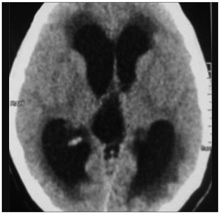
Fig. 1 CT scan brain images of first case (axial) showing hydrocephalus with dilated lateral and third ventricles.
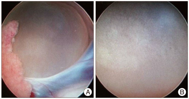
Fig. 2 A : Endoscopic view of cysticercus lesion seen obstructing the foramen of Monroe with surrounding relations. B : Cysticercus lesion appearing as the "Full moon" in the endoscopic view.
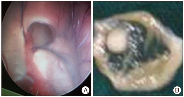
Fig. 3 A : Image showing the endoscopic view of foramen Monroe and third ventricle after the cyst removal. B : Image showing the thin walled cyst containing cysticercus larva in form of a nodule inside.
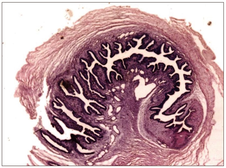
Fig. 4 Haematoxylin and eosin stained low power field image showing cysticercus larva. Cyst cavity is lined by three layers : cuticle, cellular and inner loose layer. Cephalic end of the larva showing an invaginated scolex having a sucker and hooklets is identified. Caudal end of scolex larva showing duct like invaginations surrounding a coelomic cavity.
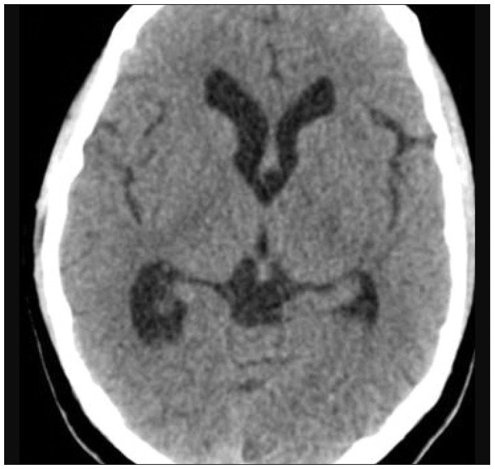
Fig. 5 Six months post operative CT scan image of the brain showing resolved hydrocephalus.
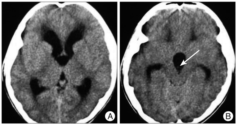
Fig. 6 Serial axial CT scan images (A) of second case showing hydrocephalus and a hyperdense nodular lesion in the third ventricle (B) (arrow).
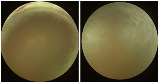
Fig. 7 Intraoperative endoscopic image showing cysticercus lesion completely obstructing foramen of Monroe with fornix seen superiorly (full moon endoscopic sign).
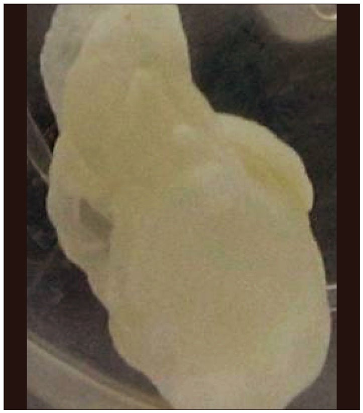
Fig. 8 Image of the cysticercus lesion removed endoscopically.
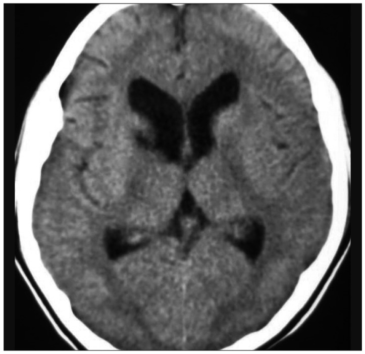
Fig. 9 Three months post operative CT brain image showing resolved hydrocephalus.
Reference
-
1. Anandh B, Mohanty A, Sampath S, Praharaj SS, Kolluri S. Endoscopic approach to intraventricular cysticercal lesions. Minim Invasive Neurosurg. 2001; 44:194–196. PMID: 11830776.
Article2. Apuzzo ML, Dobkin WR, Zee CS, Chan JC, Giannotta SL, Weiss MH. Surgical considerations in treatment of intraventricular cysticercosis. An analysis of 45 cases. J Neurosurg. 1984; 60:400–407. PMID: 6607328.3. Bergsneider M, Holly LT, Lee JH, King WA, Frazee JG. Endoscopic management of cysticercal cysts within the lateral and third ventricles. J Neurosurg. 2000; 92:14–23. PMID: 10616077.
Article4. Buxton N, Punt J. Cerebral infarction after neuroendoscopic third ventriculostomy : case report. Neurosurgery. 2000; 46:999–1001. discussion 1001-1002. PMID: 10764279.
Article5. Carpio A. Neurocysticercosis : an update. Lancet Infect Dis. 2002; 2:751–762. PMID: 12467692.6. Colli BO, Martelli N, Assirati JA Jr, Machado HR, de Vergueiro Forjaz S. Results of surgical treatment of neurocysticercosis in 69 cases. J Neurosurg. 1986; 65:309–315. PMID: 3734881.
Article7. Goel RK, Ahmad FU, Vellimana AK, Suri A, Chandra PS, Kumar R, et al. Endoscopic management of intraventricular neurocysticercosis. J Clin Neurosci. 2008; 15:1096–1101. PMID: 18653345.
Article8. Husain M, Jha DK, Rastogi M, Husain N, Gupta RK. Neuro-endoscopic management of intraventricular neurocysticercosis (NCC). Acta Neurochir (Wien). 2007; 149:341–346. PMID: 17342378.
Article9. Husain M, Rastogi M, Jha DK, Husain N, Gupta RK. Endoscopic transaqueductal removal of fourth ventricular neurocysticercosis with an angiographic catheter. Neurosurgery. 2007; 60(4 Suppl 2):249–253. discussion 254. PMID: 17415160.
Article10. Matushita H, Pinto FC, Cardeal DD, Teixeira MJ. Hydrocephalus in neurocysticercosis. Childs Nerv Syst. 2011; 27:1709–1721. PMID: 21928035.
Article11. McLaughlin MR, Wahlig JB, Kaufmann AM, Albright AL. Traumatic basilar aneurysm after endoscopic third ventriculostomy : case report. Neurosurgery. 1997; 41:1400–1403. discussion 1403-1404. PMID: 9402593.
Article12. Medina MT, DeGiorgio C. Introduction to Neurocysticercosis : a worldwide epidemic. Neurosurg Focus. 2002; 12:1–6.13. Proaño JV, Torres-Corzo J, Rodríguez-DellaVecchia R, Guizar-Sahagun G, Rangel-Castilla L. Intraventricular and subarachnoid basal cisterns neurocysticercosis : a comparative study between traditional treatment versus neuroendoscopic surgery. Childs Nerv Syst. 2009; 25:1467–1475. PMID: 19557421.
Article14. Ramos-Zúñiga R, de La Cruz-Ramírez J, Casillas-Espinosa PM, Sánchez-Prieto JA, López-Hernández MD. "Full moon" endoscopic sign in intraventricular neurocysticercosis. Minim Invasive Neurosurg. 2011; 54:90–94. PMID: 21656445.
Article15. Singh S, Gibikote SV, Shyamkumar NK. Isolated fourth ventricular cysticercus cyst : MR imaging in 4 cases with short literature review. Neurol India. 2003; 51:394–396. PMID: 14652451.16. Sinha S, Sharma BS. Intraventricular neurocysticercosis : a review of current status and management issues. Br J Neurosurg. 2012; 26:305–309. PMID: 22168964.
Article17. Suri A, Goel RK, Ahmad FU, Vellimana AK, Sharma BS, Mahapatra AK. Endoscopic excision of intraventricular neurocysticercosis in children : a series of six cases and review. Childs Nerv Syst. 2008; 24:281–285. PMID: 17994242.
Article
- Full Text Links
-
- Actions
-
Cited
- CITED
-
- Close
- Share
-
- Similar articles
-
- Transaqueductal Migration of the Neurocysticercus Cyst: Two Case Report
- Stereotactic Surgery of Neurocysticercosis
- Acquired Chiari-malformation after Ventriculoperitoneal Shunt for Hydrocepalus Associated with Neurocysticercosis
- Extraparenchymal (Racemose) Neurocysticercosis and Its Multitude Manifestations: A Comprehensive Review
- Calcified Neurocysticercosis that Invaded the Subarachnoid Space Presenting as Focal Status Epilepticus
