J Korean Neurosurg Soc.
2014 Jan;55(1):40-42. 10.3340/jkns.2014.55.1.40.
Two Cystic Cavernous Angiomas after Radiotherapy for Atypical Meningioma in Adult Woman : Case Report and Literature Review
- Affiliations
-
- 1Department of Neurological Sciences-Neurosurgery, Sapienza University of Rome, Rome, Italy. padonnarumma@hotmail.it
- KMID: 2191039
- DOI: http://doi.org/10.3340/jkns.2014.55.1.40
Abstract
- A correlation between radiation therapy and cavernoma has been suspected since 1994. Since then, only a few cases of radio-induced cavernomas have been reported in the literature (85 patients). Most of them were children, and the most frequent original tumour had been medulloblastoma. The authors report a case of two cystic cavernous angiomas after radiation therapy for atypical meningioma in adult woman. This is the first case of cavernous angioma after radiotherapy for low grade meningioma. A 39-year-old, Latin american woman was operated on for a frontal atypical meningioma with intradiploic component and adjuvant radiotherapy was delivered (6000 cGy local brain irradiation, fractionated over 6 weeks). Follow-up MR imaging showed no recurrences of the tumour and no other lesions. Ten years later, at the age of 49, she consulted for progressive drug-resistant headache. MR imaging revealed two new well defined areas of different signal intensity at the surface of each frontal pole. Both lesions were surgically removed; the histopathological diagnosis was cavernous angioma. This is the first case of cavernous angioma after radiation therapy for atypical meningioma : it confirms the development of these lesions after standard radiation therapy also in patients previously affected by non-malignant tumours.
Keyword
MeSH Terms
Figure
-
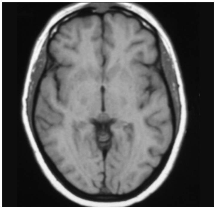
Fig. 1 Atypical menigioma. Axial T1-weighted MRI.
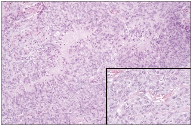
Fig. 2 Atypical menigioma. Histological examination.
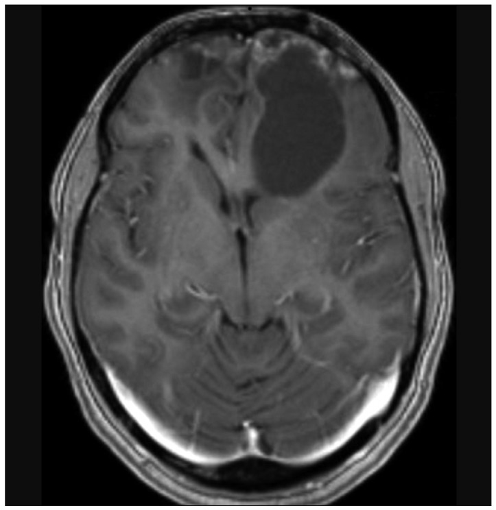
Fig. 3 Cystic cavernous angiomas. Axial gadolinium enhanced T1-weighted MRI.
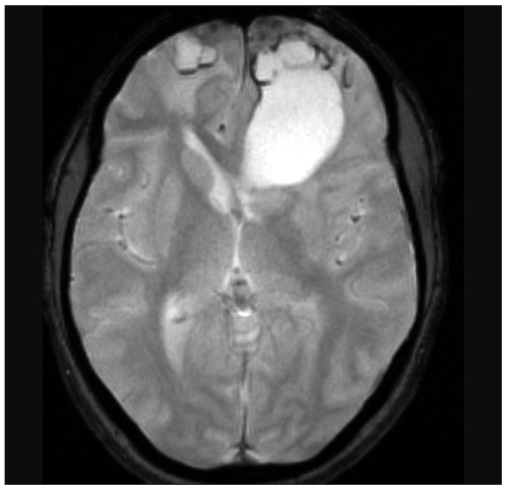
Fig. 4 Cystic cavernous angiomas. Axial GRE T2-weighted MRI.
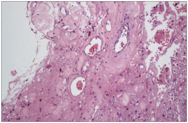
Fig. 5 Cystic cavernous angiomas. Histological examination.
Reference
-
1. Battaglia F, Uro-Coste E, Delisle MB, Tannier C. [Radiation-induced cavernoma : two cases]. Rev Neurol (Paris). 2008; 164:468–471. PMID: 18555880.2. Burn S, Gunny R, Phipps K, Gaze M, Hayward R. Incidence of cavernoma development in children after radiotherapy for brain tumors. J Neurosurg. 2007; 106(5 Suppl):379–383. PMID: 17566205.
Article3. Ciricillo SF, Cogen PH, Edwards MS. Pediatric cryptic vascular malformations : presentation, diagnosis and treatment. Pediatr Neurosurg. 1994; 20:137–147. PMID: 8161487.
Article4. Duhem R, Vinchon M, Leblond P, Soto-Ares G, Dhellemmes P. Cavernous malformations after cerebral irradiation during childhood : report of nine cases. Childs Nerv Syst. 2005; 21:922–925. PMID: 15662523.
Article5. Furuse M, Miyatake SI, Kuroiwa T. Cavernous malformation after radiation therapy for astrocytoma in adult patients : report of 2 cases. Acta Neurochir (Wien). 2005; 147:1097–1101. discussion 1101. PMID: 16021386.
Article6. Günel M, Awad IA, Finberg K, Steinberg GK, Craig HD, Cepeda O, et al. Genetic heterogeneity of inherited cerebral cavernous malformation. Neurosurgery. 1996; 38:1265–1271. PMID: 8727164.
Article7. Heckl S, Aschoff A, Kunze S. Radiation-induced cavernous hemangiomas of the brain : a late effect predominantly in children. Cancer. 2002; 94:3285–3291. PMID: 12115362.
Article8. Jain R, Robertson PL, Gandhi D, Gujar SK, Muraszko KM, Gebarski S. Radiation-induced cavernomas of the brain. AJNR Am J Neuroradiol. 2005; 26:1158–1162. PMID: 15891176.9. Keezer MR, Del Maestro R. Radiation-induced cavernous hemangiomas : case report and literature review. Can J Neurol Sci. 2009; 36:303–310. PMID: 19534329.
Article10. Moriarity JL, Wetzel M, Clatterbuck RE, Javedan S, Sheppard JM, Hoenig-Rigamonti K, et al. The natural history of cavernous malformations : a prospective study of 68 patients. Neurosurgery. 1999; 44:1166–1171. discussion 1172-1173. PMID: 10371615.
Article11. Nimjee SM, Powers CJ, Bulsara KR. Review of the literature on de novo formation of cavernous malformations of the central nervous system after radiation therapy. Neurosurg Focus. 2006; 21:e4. PMID: 16859257.
Article12. Nyáry I, Major O, Hanzély Z, Szeifert GT. Histopathological findings in a surgically resected thalamic cavernous hemangioma 1 year after 40-Gy irradiation. J Neurosurg. 2005; 102(Suppl):56–58. PMID: 15662782.
Article13. Rigamonti D, Hadley MN, Drayer BP, Johnson PC, Hoenig-Rigamonti K, Knight JT, et al. Cerebral cavernous malformations. Incidence and familial occurrence. N Engl J Med. 1988; 319:343–347. PMID: 3393196.14. Tomlinson FH, Houser OW, Scheithauer BW, Sundt TM Jr, Okazaki H, Parisi JE. Angiographically occult vascular malformations : a correlative study of features on magnetic resonance imaging and histological examination. Neurosurgery. 1994; 34:792–799. discussion 799-800. PMID: 8052376.
