Primary Eosinophilic Granuloma of Adult Cervical Spine Presenting as a Radiculomyelopathy
- Affiliations
-
- 1Department of Neurosurgery, Kyungpook National University Hospital, Daegu, Korea. nskimkt7@gmail.com
- KMID: 2190860
- DOI: http://doi.org/10.3340/jkns.2013.54.1.54
Abstract
- We report a case of 29-year-old man diagnosed as a primary eosinophilic granuloma (EG) lesion of the seventh cervical vertebra. He had paresthesia on both arms, and grasping weakness for 10 days. Cervical magnetic resonance image (MRI) showed an enhancing mass with ventral epidural bulging and cord compression on the seventh cervical vertebra. Additionally, we performed spine series MRI, bone scan and positive emission tomography for confirmation of other bone lesions. These studies showed no other pathological lesions. He underwent anterior cervical corpectomy of the seventh cervical vertebra and plate fixation with iliac bone graft. After surgical management, neurological symptoms were much improved. Histopathologic evaluation confirmed the diagnosis of EG. There was no evidence of tumor recurrence at 12 months postoperative cervical MRI follow-up. We reported symptomatic primary EG of cervical spine successfully treated with surgical resection.
MeSH Terms
Figure
-

Fig. 1 Preoperative radiographic images show tumorous condition. MRI showed collapse of C7 vertebral body and extension to paravertebral soft tissue and epidural space without invasion to adjacent disc spaces. In addition, epidural extension produced cord compression without definite cord signal change. Computed tomography scan and simple radiograph revealed severe bony destruction and sparing of the posterior column. A : T1 weighted sagittal MRI scan. B : T2 weighted sagittal MRI scan. C : T1-weighted gadolinium-enhanced axial MRI scan. D : Simple lateral radiographs at presentation. E : Coronal CT scan. F : Axial CT scan.
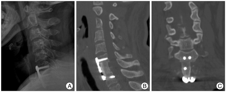
Fig. 2 POD 1 radiographic images show C7 corpectomy state. A : Simple lateral radiograph. B : Cervical sagittal CT scan. C : Cervical coronal CT scan.

Fig. 3 Twelve month follow up MRI demonstrate no evidence of recurrence. A : T2 weight sagittal MRI scan. B : T1 weight sagittal MRI scan.
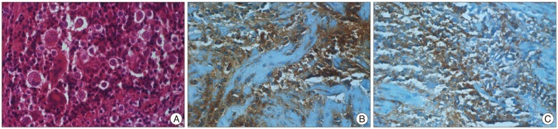
Fig. 4 Histopathological examination of the patient. A : On hematoxylin-eosin staining (magnification ×400), the section consists of many cells showing that Langerhans cell features have convoluted nuclei and linear grooves. This lesion was associated with numerous giant cells and eosinophils. B and C : Immunostains for S100 (Magnification ×400) (B) and CD1a (magnification ×400) (C) show diffuse strong cytoplasmic staining on the Largerhans cells.
Reference
-
1. Azouz EM, Saigal G, Rodriguez MM, Podda A. Langerhans' cell histiocytosis : pathology, imaging and treatment of skeletal involvement. Pediatr Radiol. 2005; 35:103–115. PMID: 15289942.
Article2. Bertram C, Madert J, Eggers C. Eosinophilic granuloma of the cervical spine. Spine (Phila Pa 1976). 2002; 27:1408–1413. PMID: 12131737.
Article3. Brown CW, Jarvis JG, Letts M, Carpenter B. Treatment and outcome of vertebral Langerhans cell histiocytosis at the Children's Hospital of Eastern Ontario. Can J Surg. 2005; 48:230–236. PMID: 16013628.4. Bunch WH. Orthopedic and rehabilitation aspects of eosinophilic granuloma. Am J Pediatr Hematol Oncol. 1981; 3:151–156. PMID: 7304858.
Article5. Glotzbecker MP, Carpentieri DF, Dormans JP. Langerhans cell histiocytosis : a primary viral infection of bone? Human herpes virus 6 latent protein detected in lymphocytes from tissue of children. J Pediatr Orthop. 2004; 24:123–129. PMID: 14676546.6. Howarth DM, Gilchrist GS, Mullan BP, Wiseman GA, Edmonson JH, Schomberg PJ. Langerhans cell histiocytosis : diagnosis, natural history, management, and outcome. Cancer. 1999; 85:2278–2290. PMID: 10326709.7. Huang W, Yang X, Cao D, Xiao J, Yang M, Feng D, et al. Eosinophilic granuloma of spine in adults : a report of 30 cases and outcome. Acta Neurochir (Wien). 2010; 152:1129–1137. PMID: 20396916.
Article8. Islinger RB, Kuklo TR, Owens BD, Horan PJ, Choma TJ, Murphey MD, et al. Langerhans' cell histiocytosis in patients older than 21 years. Clin Orthop Relat Res. 2000; 231–235. PMID: 11039811.
Article9. Jang KS, Jung YY, Kim SW. Langerhans cell histiocytosis causing cervical myelopathy in a child. J Korean Neurosurg Soc. 2010; 47:458–460. PMID: 20617093.
Article10. Jiang L, Liu ZJ, Liu XG, Zhong WQ, Ma QJ, Wei F, et al. Langerhans cell histiocytosis of the cervical spine : a single Chinese institution experience with thirty cases. Spine (Phila Pa 1976). 2010; 35:E8–E15. PMID: 20042947.11. Kilborn TN, Teh J, Goodman TR. Paediatric manifestations of Langerhans cell histiocytosis : a review of the clinical and radiological findings. Clin Radiol. 2003; 58:269–278. PMID: 12662947.
Article12. Lichtenstein L. Histiocytosis X; integration of eosinophilic granuloma of bone, Letterer-Siwe disease, and Schüller-Christian disease as related manifestations of a single nosologic entity. AMA Arch Pathol. 1953; 56:84–102. PMID: 13057466.13. Peng XS, Pan T, Chen LY, Huang G, Wang J. Langerhans' cell histiocytosis of the spine in children with soft tissue extension and chemotherapy. Int Orthop. 2009; 33:731–736. PMID: 18338168.
Article14. Richter MP, D'Angio GJ. The role of radiation therapy in the management of children with histiocytosis X. Am J Pediatr Hematol Oncol. 1981; 3:161–163. PMID: 7030100.
Article15. Sayhan S, Altinel D, Erguden C, Kizmazoglu C, Guray M, Acar U. Langerhans cell histiocytosis of the cervical spine in an adult : a case report. Turk Neurosurg. 2010; 20:409–412. PMID: 20669118.16. Scarpinati M, Artico M, Artizzu S. Spinal cord compression by eosinophilic granuloma of the cervical spine. Case report and review of the literature. Neurosurg Rev. 1995; 18:209–212. PMID: 8570070.
Article17. Seimon LP. Eosinophil granuloma of the spine. J Pediatr Orthop. 1981; 1:371–376. PMID: 7334115.
Article18. Simanski C, Bouillon B, Brockmann M, Tiling T. The Langerhans' cell histiocytosis (eosinophilic granuloma) of the cervical spine: a rare diagnosis of cervical pain. Magn Reson Imaging. 2004; 22:589–594. PMID: 15120180.
Article19. Tan G, Samson I, De Wever I, Goffin J, Demaerel P, Van Gool SW. Langerhans cell histiocytosis of the cervical spine : a single institution experience in four patients. J Pediatr Orthop B. 2004; 13:123–126. PMID: 15076592.
Article20. Vadivelu S, Mangano FT, Miller CR, Leonard JR. Multifocal Langerhans cell histiocytosis of the pediatric spine : a case report and literature review. Childs Nerv Syst. 2007; 23:127–131. PMID: 17021733.
Article21. Wei MA, Ruixue MA. Solitary spinal eosinophilic granuloma in children. J Pediatr Orthop B. 2006; 15:316–319. PMID: 16891956.
Article22. Yeom JS, Lee CK, Shin HY, Lee CS, Han CS, Chang H. Langerhans' cell histiocytosis of the spine. Analysis of twenty-three cases. Spine (Phila Pa 1976). 1999; 24:1740–1749. PMID: 10472109.
