Measurement of Critical Structures around Paraclinoidal Area : A Cadaveric Morphometric Study
- Affiliations
-
- 1Department of Neurosurgery, College of Medicine, Dong-A University, Busan, Korea. ns2000@dau.ac.kr
- 2Department of Anatomy, College of Medicine, Dong-A University, Busan, Korea.
- KMID: 2190851
- DOI: http://doi.org/10.3340/jkns.2013.54.1.14
Abstract
OBJECTIVE
Although removal of the anterior clinoid process (ACP) is essential surgical technique, studies about quantitative measurements of the space broadening by the anterior clinoidectomy are rare. The purposes of this study are to investigate the dimension of the ACP, to quantify the improved exposure of the parasellar space after extradural anterior clinoidectomy and to measure the correlation of each structure around the paraclinoidal area.
METHODS
Eleven formalin-fixed Korean adult cadaveric heads were used and frontotemporal craniotomies were done bilaterally. The length of C6 segment of the internal carotid artery on its lateral and medial side and optic nerve length were checked before and after anterior clinoidectomy. The basal width and height of the ACP were measured. The relationships among the paraclinoidal structures were assessed. The origin and projection of the ophthalmic artery (OA) were investigated.
RESULTS
The mean values of intradural basal width and height of the ACP were 10.82 mm and 7.61 mm respectively. The mean length of the C6 lateral and medial side increased 49%. The mean length of optic nerve increased 97%. At the parasellar area, the lengths from the optic strut to the falciform liament, distal dural ring, origin of OA were 6.69 mm, 9.36 mm and 5.99 mm, respectively. The distance between CN III and IV was 11.06 mm.
CONCLUSION
With the removal of ACP, exposure of the C6 segments and optic nerve can expand 49% and 97%, respectively. This technique should be among a surgeon's essential skills for treating lesions around the parasellar area.
Keyword
Figure
-
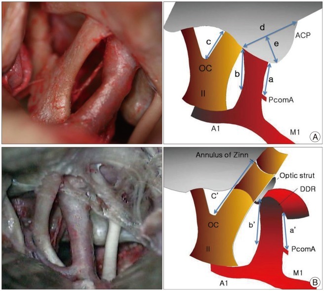
Fig. 1 A: Measurements before anterior clinoidectomy. Exposure of the ICA and optic nerve in operative view (left) and schematic image (right). (a) and (b) represent the lengths of the exposed ICA on its lateral and medial sides, respectively. (c) is the optic nerve length from the chiasm to the falciform ligament, (d) and (e) are the intradural basal width and height of the ACP, respectively. B: Exposure of the ICA and optic nerve after anterior clinoidectomy in operative view (left) and schematic image (right). (a') and (b') represent the extended ICA length on its lateral and medial sides, respectively. They correspond to the entire C6 segment of the ICA. (c') is the optic nerve length from the chiasm to the annulus of Zinn. ACP: anterior clinoid process, OC: optic chiasm, PcomA: posterior communicating artery, ICA: internal carotid artery, DDR: distal dural ring, II: optic nerve, A1: anterior cerebral artery, M1: middle cerebral artery.
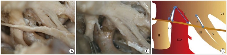
Fig. 2 Operative views (A and B) and a schematic iamge (C) after extradural anterior clinoidectomy. A: Before releasing the DDR and opening the optic sheath. The distance between the optic strut and the falciform ligament was checked (f). The distance between the lateral portion of the DDR and the optic strut was checked (g). The distance between the entry of the CN III into the cavernous sinus and the point of intersection with the CN IV was measured (h). B: Operative view after opening of the optic sheath, and releasing of the DDR. The OA was exposed and the distance between its origin and the optic strut is checked (i). DDR: distal dural ring, OA: ophthalmic artery, ICA: internal carotid artery, II: optic nerve, III: oculomotor nerve, IV: trochlear nerve, V1: ophthalmic branch of trigeminal nerve.
Reference
-
1. Andaluz N, Beretta F, Bernucci C, Keller JT, Zuccarello M. Evidence for the improved exposure of the ophthalmic segment of the internal carotid artery after anterior clinoidectomy: morphometric analysis. Acta Neurochir (Wien). 2006; 148:971–975. discussion 975-976. PMID: 16917665.
Article2. Bouthillier A, van Loveren HR, Keller JT. Segments of the internal carotid artery: a new classification. Neurosurgery. 1996; 38:425–432. discussion 432-433. PMID: 8837792.
Article3. Chang DJ. The "no-drill" technique of anterior clinoidectomy: a cranial base approach to the paraclinoid and parasellar region. Neurosurgery. 2009; 64(3 Suppl):ons96–ons105. discussion ons105-106. PMID: 19240577.
Article4. Collignon F, Link M. Paraclinoid and cavernous sinus regions: measurement of critical structures relevant for surgical procedure. Clin Anat. 2005; 18:3–9. PMID: 15597376.
Article5. Coscarella E, Başkaya MK, Morcos JJ. An alternative extradural exposure to the anterior clinoid process: the superior orbital fissure as a surgical corridor. Neurosurgery. 2003; 53:162–166. discussion 166-167. PMID: 12823885.
Article6. De Jesús O. The clinoidal space: anatomical review and surgical implications. Acta Neurochir (Wien). 1997; 139:361–365. PMID: 9202779.
Article7. Dolenc VV. A combined epi- and subdural direct approach to carotid-ophthalmic artery aneurysms. J Neurosurg. 1985; 62:667–672. PMID: 3989589.
Article8. Erdogmus S, Govsa F. Anatomic features of the intracranial and intracanalicular portions of ophthalmic artery: for the surgical procedures. Neurosurg Rev. 2006; 29:213–218. PMID: 16775743.
Article9. Froelich SC, Aziz KM, Levine NB, Theodosopoulos PV, van Loveren HR, Keller JT. Refinement of the extradural anterior clinoidectomy: surgical anatomy of the orbitotemporal periosteal fold. Neurosurgery. 2007; 61(5 Suppl 2):179–185. discussion 185-186. PMID: 18091231.10. Gibo H, Lenkey C, Rhoton AL Jr. Microsurgical anatomy of the supraclinoid portion of the internal carotid artery. J Neurosurg. 1981; 55:560–574. PMID: 7277004.
Article11. Huynh-Le P, Natori Y, Sasaki T. Surgical anatomy of the anterior clinoid process. J Clin Neurosci. 2004; 11:283–287. PMID: 14975418.
Article12. Hwang YS, Park SK, Shin HS, Kim SJ, Lee JH, Evans J. Pre -vs. post-anterior clinoidectomy measurements of the optic nerve, internal carotid artery, and optico-carotid triangle: a cadaveric morphometric study. J Korean Neurosurg Soc. 1999; 28:1082–1088.13. Inoue T, Rhoton AL Jr, Theele D, Barry ME. Surgical approaches to the cavernous sinus: a microsurgical study. Neurosurgery. 1990; 26:903–932. PMID: 2362670.
Article14. Kulwin C, Tubbs RS, Cohen-Gadol AA. Anterior clinoidectomy: description of an alternative hybrid method and a review of the current techniques with an emphasis on complication avoidance. Surg Neurol Int. 2011; 2:140. PMID: 22059135.
Article15. Lee HY, Chung IH, Choi BY, Lee KS. Anterior clinoid process and optic strut in Koreans. Yonsei Med J. 1997; 38:151–154. PMID: 9259614.
Article16. Mori K, Yamamoto T, Nakao Y, Esaki T. Surgical Simulation of Extradural Anterior Clinoidectomy through the Trans-superior Orbital Fissure Approach Using a Dissectable Three-dimensional Skull Base Model with Artificial Cavernous Sinus. Skull Base. 2010; 20:229–236. PMID: 21311615.
Article17. Noguchi A, Balasingam V, Shiokawa Y, McMenomey SO, Delashaw JB Jr. Extradural anterior clinoidectomy. Technical note. J Neurosurg. 2005; 102:945–950. PMID: 15926728.18. Romani R, Elsharkawy A, Laakso A, Kangasniemi M, Hernesniemi J. Complications of anterior clinoidectomy through lateral supraorbital approach. World Neurosurg. 2012; 77:698–703. PMID: 22120307.
Article19. Takahashi JA, Kawarazaki A, Hashimoto N. Intradural en-bloc removal of the anterior clinoid process. Acta Neurochir (Wien). 2004; 146:505–509. PMID: 15118888.
Article
- Full Text Links
-
- Actions
-
Cited
- CITED
-
- Close
- Share
-
- Similar articles
-
- Morphometric Study of the Nerve Roots Around the Lateral Mass for Posterior Foraminotomy
- Morphometric study of fossa ovale in human cadaveric hearts: embryological and clinical relevance
- Comparison of the Results between Cadaveric and Radiological Measurements of Calcaneus
- Morphometric Analysis of Malignant Lymphoma
- The Relationship between Disc Degeneration and Morphologic Changes in the Intervertebral Foramen of the Cervical Spine: A Cadaveric MRI and CT Study
