Morphometric study of fossa ovale in human cadaveric hearts: embryological and clinical relevance
- Affiliations
-
- 1Department of Anatomy, ESIC Medical College & Hospital, Hyderabad, India
- 2Department of Anatomy, All India Institute of Medical Sciences, Bibinagar, Hyderabad, India
- KMID: 2514582
- DOI: http://doi.org/10.5115/acb.20.286
Abstract
- Atrial septal defect (ASD) is the 5th common congenital abnormality at birth. Secundum atrial defect and patent foramen ovale (PFO) are the most common atrial septal defects. In this setting, the anatomical functional characterization of the interatrial septum seems to be of paramount importance not only for device selection but also for therapeutic intervention. This study was carried out to evaluate the morphometric parameters of fossa ovale (FOv) in the human adult cadaveric hearts. For this study, 50 normal cadaveric human hearts available in the department of Anatomy over the period of 3 years were used where size, position, shape, nature of the FOv was noted. The size of the fossa was measured and prominence, location, and extent of the limbus fossa ovalis were observed. The probe patency of foramen ovale (FO) was confirmed. In most specimens, the fossa was oval (80%), the average transverse diameter was 24.21 mm, and the vertical diameter 26.84 mm. In 84% rim was raised. In 56% of cases, the fossa was present at the middle of the interatrial septum. The patency of foramen was observed in 3%. The findings of the present study provide pertinent information on the morphology of the FOv, which may be useful for device selection in treating ASDs and PFO. This would definitely help the clinicians in a deeper understanding of the region as very few cadaveric studies are available in the literature at present.
Figure
-
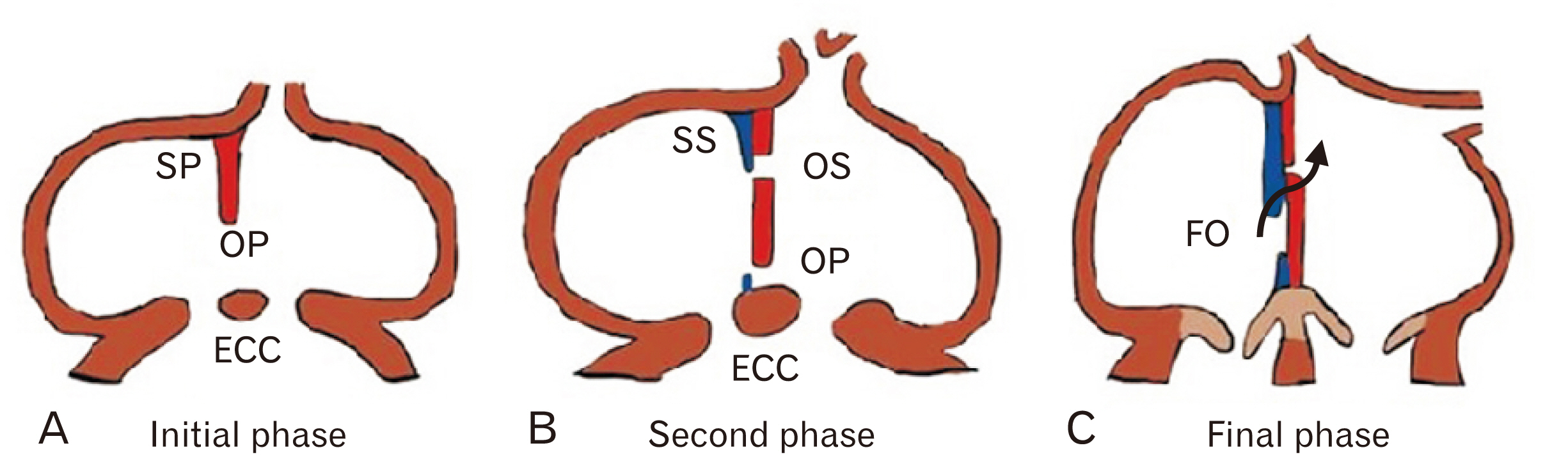
Fig. 1 Normal formation of IAS. (A) SP a sickle-shaped crest that descends from the roof of the primitive atrium. Opening between SP and ECC is OP. (B) SP fusing with ECC, perforation in the upper part of SP forms OS. New fold appears from roof as SS. (C) Opening between lower end of SS and upper end of SP is FO. ECC, endocardial cushion; FO, foramen ovale; IAS, inter atrial septum; OP, ostium primum; OS, ostium secundum; SP, septum primum; SS, septum secundum.
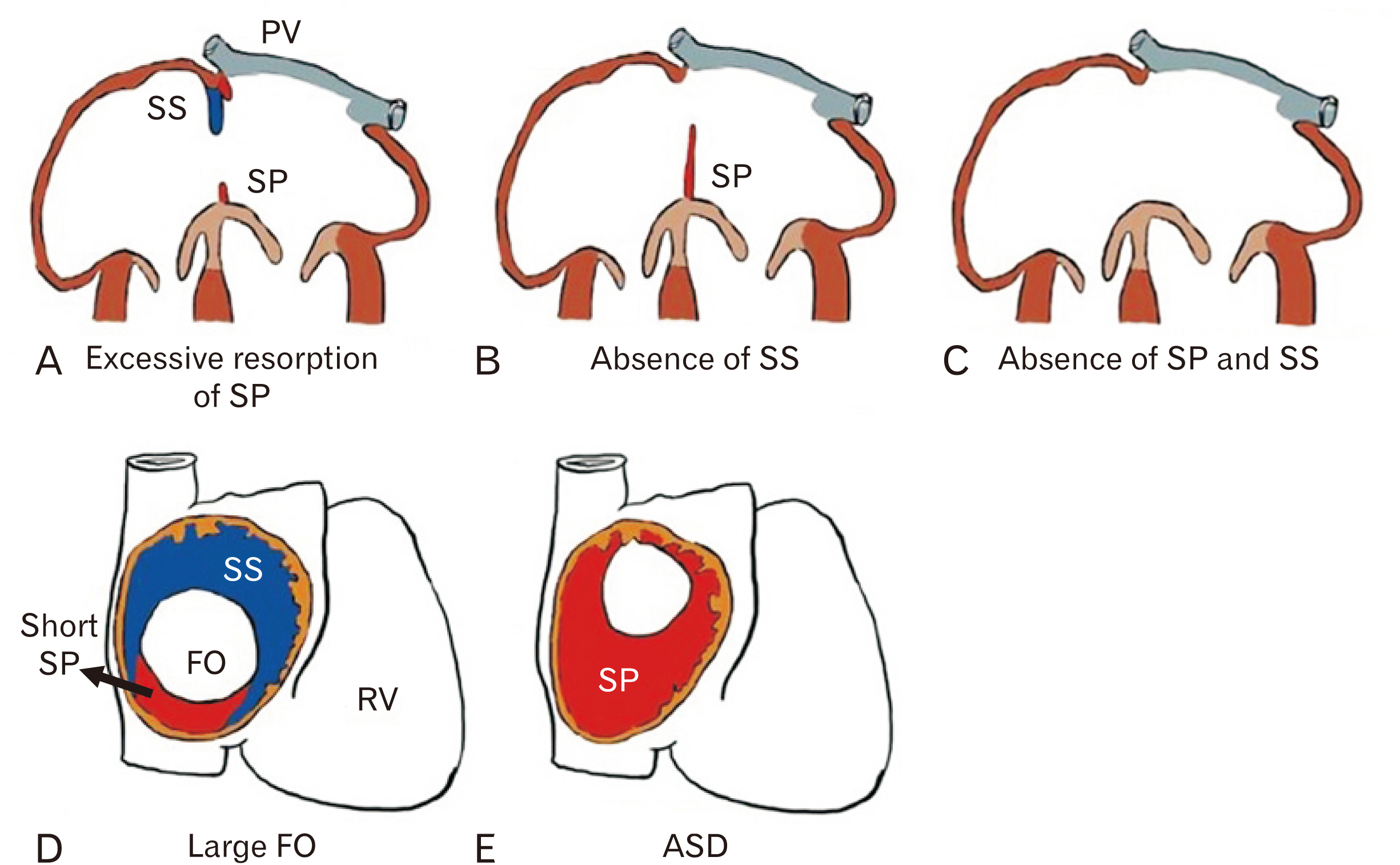
Fig. 2 Developmental defects of IAS formation showing. (A) Excessive resorption of SP. (B): Absence of SS. (C) Absence of both SP and SS, leading to a common atrium or Cor triloculare biventriculare. (D) Showing large FO. (E) Showing ASD. ASD, atrial septal defect; IAS, inter atrial septum;O, foramen ovale; PV, pulmonary veins; RV, right ventricle; SP, septum primum; SS, septum secundum.
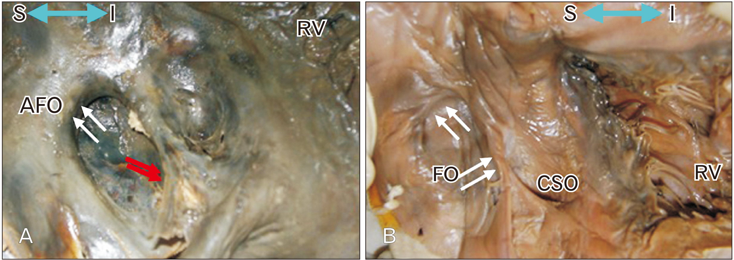
Fig. 3 Elliptical shape of FOv. (A) Showing raised margin in whole circumference AFO. In upper part shows slit like recess or pouch (white arrows). In lower part a deep recess present (red arrows). (B) Raised margin is present only in upper and anterior part of FO with slit like recess or pouch (white arrows). Blue double headed arrow showing S and I view. AFO, annulus fossa ovalis; CSO, coronary sinus opening; FO, foramen ovale; FOv, fossa ovale; RV, right ventricle; I, inferior; S, superior.
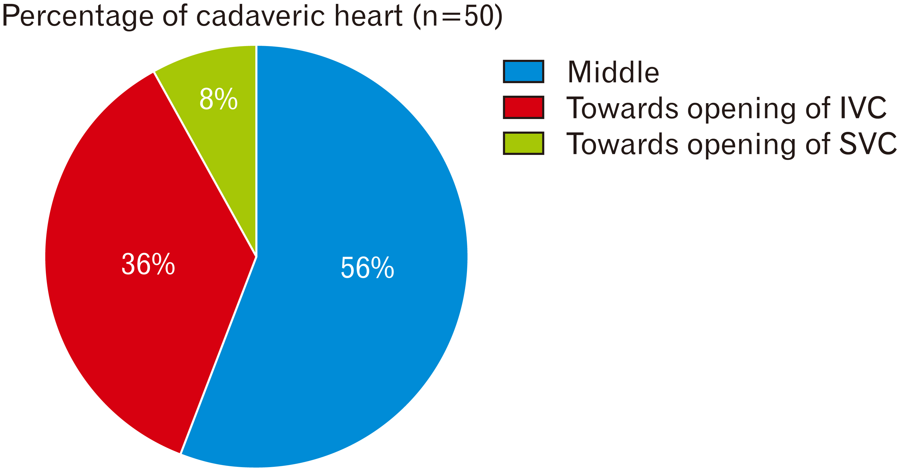
Fig. 4 Showing Position of FOv in the interatrial septum in cadaveric hearts (n=50). FOv, fossa ovale; IVC, inferior vena cava; SVC, superior vena cava.

Fig. 5 Showing different shapes of FOv in the interatrial septum in cadaveric hearts (n=50). FOv, fossa ovale.
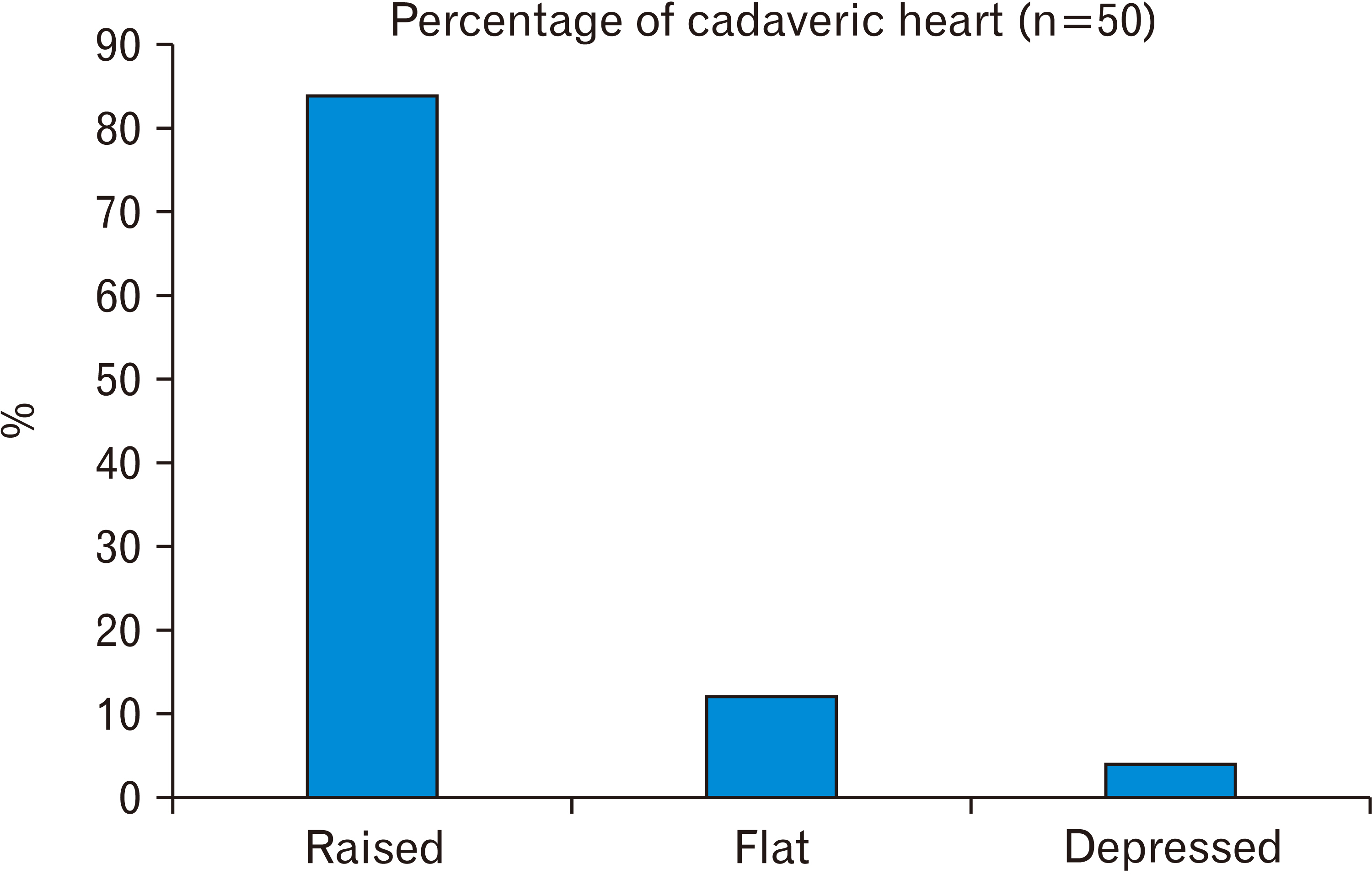
Fig. 6 Showing extent and location of Limbus fossa ovalis in the interatrial septum in cadaveric hearts (n=50).
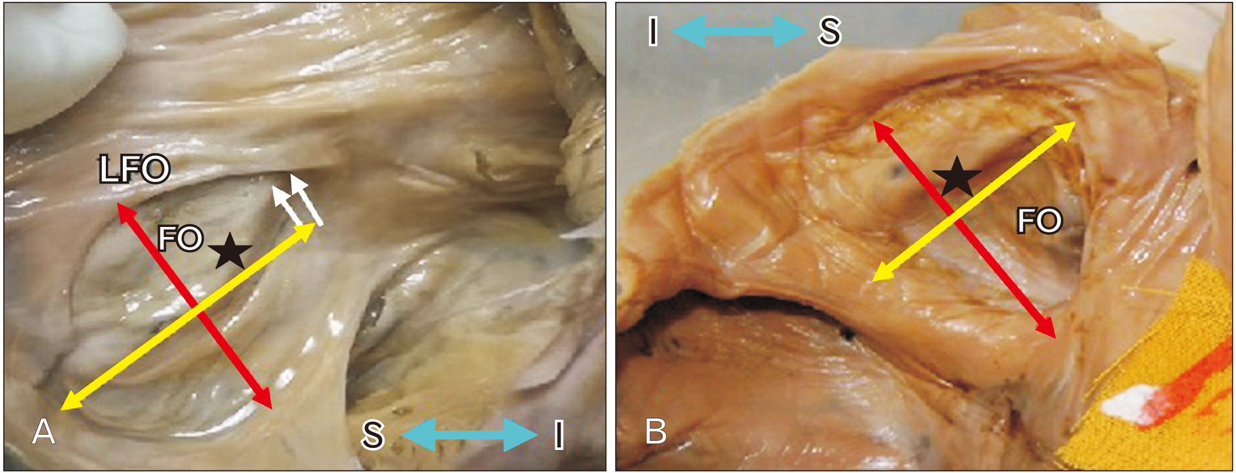
Fig. 7 Large FOv. (A) Showing deep recess in the superior margin (two white arrows), redundancy present in the middle part (asterisk). (B) Showing redundancy in the right part of FOv. In the right part showing bulging towards left atrium. Red arrow is showing vertical diameter, Yellow arrow is showing transverse diameter. Blue double headed arrow showing S and I view. FO, foramen ovale; FOv, fossa ovale; I, inferior; LFO, limbus fossa ovalis; S, superior.

Fig. 8 (A) Showing fibrous brands in the inferior part (yellow arrows). (B) Showing a membrane (red asterisk) in antero superior part of FOv. Blue double headed arrow showing S and I view. FO, foramen ovale; FOv, fossa ovale; I, inferior; S, superior.

Fig. 9 Showing shapes of FOv. (A) Oval shape, (B) Round. Red arrow is showing vertical diameter, Yellow arrow is showing transverse diameter. Double-headed red arrow indicates the long axis and yellow arrow indicates width. Blue double headed arrow showing S and I view. CSO, coronary sinus opening; FO, foramen ovale; FOv, fossa ovale; I, inferior; RV, right ventricle; S, superior; TCV, tricuspid valve.
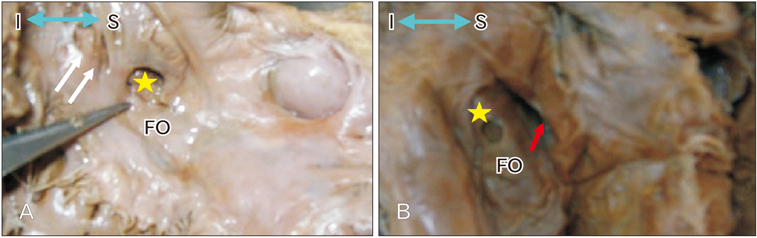
Fig. 10 Right surface of the interatrial septum. (A) Showing presence of a foramen (yellow asterik). (B) Showing deep recess (red arrow). Blue double headed arrow showing S and I view. FO, foramen ovale; I, inferior; S, superior.
Reference
-
References
1. Rosse C, Gaddum-Rosse P. 1997. Hollinshead's textbook of anatomy. 5th ed. Lippincott-Raven;Philadelphia: p. 473.2. Moore KL, Persaud TVN. Moore KL, Persaud TVN, editors. 2003. The cardiovascular system. The Developing Human: Clinically Oriented Embryology. 7th ed. Saunders;Philadelphia: p. 340–5.3. Sadler TW. Sadler TW, editor. 2012. Cardiovascular system. Langman's Medical Embryology. 12th ed. Lippincott Williams & Wilkins;Philadelphia: p. 162–200.4. Standring S, Gray H. 2008. Gray's anatomy: the anatomical basis of clinical practice. 40th ed. Churchill Livingstone/Elsevier;Edinburgh: p. 964.5. Nashat H, Montanaro C, Li W, Kempny A, Wort SJ, Dimopoulos K, Gatzoulis MA, Babu-Narayan SV. 2018; Atrial septal defects and pulmonary arterial hypertension. J Thorac Dis. 10(Suppl 24):S2953–65. DOI: 10.21037/jtd.2018.08.92. PMID: 30305956. PMCID: PMC6174141.
Article6. Schott JJ, Benson DW, Basson CT, Pease W, Silberbach GM, Moak JP, Maron BJ, Seidman CE, Seidman JG. 1998; Congenital heart disease caused by mutations in the transcription factor NKX2-5. Science. 281:108–11. DOI: 10.1126/science.281.5373.108. PMID: 9651244.
Article7. Mori AD, Bruneau BG. 2004; TBX5 mutations and congenital heart disease: Holt-Oram syndrome revealed. Curr Opin Cardiol. 19:211–5. DOI: 10.1097/00001573-200405000-00004. PMID: 15096952.
Article8. Garg V, Kathiriya IS, Barnes R, Schluterman MK, King IN, Butler CA, Rothrock CR, Eapen RS, Hirayama-Yamada K, Joo K, Matsuoka R, Cohen JC, ivastava D Sr. 2003; GATA4 mutations cause human congenital heart defects and reveal an interaction with TBX5. Nature. 424:443–7. DOI: 10.1038/nature01827. PMID: 12845333.
Article9. Davison P, Clift PF, Steeds RP. 2010; The role of echocardiography in diagnosis, monitoring closure and post-procedural assessment of patent foramen ovale. Eur J Echocardiogr. 11:i27–34. DOI: 10.1093/ejechocard/jeq120. PMID: 21078836.
Article10. Mirzaali M, Dooley M, Wynne D, Cooter N, Lee L, Haworth P, Saha R, Gainsborough N, Hildick-Smith D. 2015; Patent foramen ovale closure following cryptogenic stroke or transient ischaemic attack: long-term follow-up of 301 cases. Catheter Cardiovasc Interv. 86:1078–84. DOI: 10.1002/ccd.26080. PMID: 26105198.
Article11. Nighoghossian N, Perinetti M, Barthelet M, Adeleine P, Trouillas P. 1996; Potential cardioembolic sources of stroke in patients less than 60 years of age. Eur Heart J. 17:590–4. DOI: 10.1093/oxfordjournals.eurheartj.a014913. PMID: 8733093.
Article12. Milev I, Zafirovska P, Zimbakov Z, Idrizi S, Ampova-Sokolov V, Gorgieva E, Ilievska L, Tosheski G, Hristov N, Georgievska-Ismail L, Anguseva T, Mitrev Z. 2016; Transcatheter closure of patent foramen ovale: a single center experience. Open Access Maced J Med Sci. 4:613–8. DOI: 10.3889/oamjms.2016.113. PMID: 28028400. PMCID: PMC5175508.
Article13. Overell JR, Bone I, Lees KR. 2000; Interatrial septal abnormalities and stroke: a meta-analysis of case-control studies. Neurology. 55:1172–9. DOI: 10.1212/WNL.55.8.1172. PMID: 11071496.
Article14. Nietlispach F, Meier B. 2015; Percutaneous closure of patent foramen ovale: safe and effective but underutilized. Expert Rev Cardiovasc Ther. 13:121–3. DOI: 10.1586/14779072.2015.1000305. PMID: 25556896.
Article15. Rigatelli G, Magro B, Oliva L. 2011; Anatomo-functional characterization of interatrial septum for catheter-based interventions. Am J Cardiovasc Dis. 1:227–35. PMID: 22254200. PMCID: PMC3253514.16. Marelli AJ, Mackie AS, Ionescu-Ittu R, Rahme E, Pilote L. 2007; Congenital heart disease in the general population: changing prevalence and age distribution. Circulation. 115:163–72. DOI: 10.1161/CIRCULATIONAHA.106.627224. PMID: 17210844.17. Rigatelli G, Rigatelli G. 2005; Congenital heart diseases in aged patients: clinical features, diagnosis, and therapeutic indications based on the analysis of a twenty five-year Medline search. Cardiol Rev. 13:293–6. DOI: 10.1097/01.crd.0000145928.08280.ef. PMID: 16230886.18. Reig J, Mirapeix R, Jornet A, Petit M. 1997; Morphologic characteristics of the fossa ovalis as an anatomic basis for transseptal catheterization. Surg Radiol Anat. 19:279–82. DOI: 10.1007/BF01637589. PMID: 9413071.
Article19. Kanani SD, Tank K, Patil DS, Nirvan AB, Dave RV. 2014; Cadaveric study of fossa ovalis. BJKines-NJBAS. 6:1–4.20. Fraisse A, Latchman M, Sharma SR, Bayburt S, Amedro P, di Salvo G, Baruteau AE. 2018; Atrial septal defect closure: indications and contra-indications. J Thorac Dis. 10(Suppl 24):S2874–81. DOI: 10.21037/jtd.2018.08.111. PMID: 30305947. PMCID: PMC6174144.
Article21. Chan KC, Godman MJ. 1993; Morphological variations of fossa ovalis atrial septal defects (secundum): feasibility for transcutaneous closure with the clam-shell device. Br Heart J. 69:52–5. DOI: 10.1136/hrt.69.1.52. PMID: 8457395. PMCID: PMC1024917.
Article22. Joshi SD, Chawre HK, Joshi SS. 2016; Morphological study of fossa ovalis and its clinical relevance. Indian Heart J. 68:147–52. DOI: 10.1016/j.ihj.2015.08.001. PMID: 27133322. PMCID: PMC4867950.
Article23. Kydd AC, Das P, Hoole S, Shapiro LM, Rana BS. 2013; Defining patent foramen ovale morphology using three-dimensional transoesophageal echocardiography and relationship to shunt size. Heart. 99(Suppl S2):A72. DOI: 10.1136/heartjnl-2013-304019.119.24. Naqvi N, McCarthy KP, Ho SY. 2018; Anatomy of the atrial septum and interatrial communications. J Thorac Dis. 10(Suppl 24):S2837–47. DOI: 10.21037/jtd.2018.02.18. PMID: 30305943. PMCID: PMC6174145.
Article25. Rana BS, Shapiro LM, McCarthy KP, Ho SY. 2010; Three-dimensional imaging of the atrial septum and patent foramen ovale anatomy: defining the morphological phenotypes of patent foramen ovale. Eur J Echocardiogr. 11:i19–25. DOI: 10.1093/ejechocard/jeq122. PMID: 21078835.
Article26. Kim JS, Virágh S, Moorman AF, Anderson RH, Lamers WH. 2001; Development of the myocardium of the atrioventricular canal and the vestibular spine in the human heart. Circ Res. 88:395–402. DOI: 10.1161/01.RES.88.4.395. PMID: 11230106.
Article27. Anderson RH, Brown NA, Webb S. 2002; Development and structure of the atrial septum. Heart. 88:104–10. DOI: 10.1136/heart.88.1.104. PMID: 12067964. PMCID: PMC1767197.
Article28. Teo KS, Disney PJ, Dundon BK, Worthley MI, Brown MA, Sanders P, Worthley SG. 2010; Assessment of atrial septal defects in adults comparing cardiovascular magnetic resonance with transoesophageal echocardiography. J Cardiovasc Magn Reson. 12:44. DOI: 10.1186/1532-429X-12-44. PMID: 20663157. PMCID: PMC2912273.
Article29. Klimek-Piotrowska W, Hołda MK, Koziej M, Piątek K, Hołda J. 2016; Anatomy of the true interatrial septum for transseptal access to the left atrium. Ann Anat. 205:60–4. DOI: 10.1016/j.aanat.2016.01.009. PMID: 26879344.
Article30. Bhatnagar KP, Nettleton GS, Campbell FR, Wagner CE, Kuwabara N, Muresian H. 2006; Chiari anomalies in the human right atrium. Clin Anat. 19:510–6. DOI: 10.1002/ca.20195. PMID: 16258973.
Article31. Krishnan SC, Salazar M. 2010; Septal pouch in the left atrium: a new anatomical entity with potential for embolic complications. JACC Cardiovasc Interv. 3:98–104. DOI: 10.1016/j.jcin.2009.07.017. PMID: 20129577.
- Full Text Links
-
- Actions
-
Cited
- CITED
-
- Close
- Share
-
- Similar articles
-
- Morphometric Study of the Extradural Middle Cranial Fossa for Transpetrosal Surgery
- Anatomical and radiological angiographic study of the coronary ostia in the adult human hearts and their clinical significance
- Unusual Course of the Accessory Meningeal Artery
- High Resolution CT Evaluation on the Morphologic Characteristics and Variations of Foramen Ovale and Adjacent Foramina in the Skull Base
- Evaluation of morphometric features of fossa navicularis using cone-beam computed tomography in a Turkish subpopulation
