J Korean Assoc Oral Maxillofac Surg.
2011 Feb;37(1):43-48. 10.5125/jkaoms.2011.37.1.43.
An anatomical study on the mandibular medial surface by CBCT analysis for safer implant placement
- Affiliations
-
- 1Department of Oral and Maxillofacial Surgery, School of Dentistry, Kyung-Hee University, Seoul, Korea. kyukab@khu.ac.kr
- KMID: 2189805
- DOI: http://doi.org/10.5125/jkaoms.2011.37.1.43
Abstract
- INTRODUCTION
This study examined the anatomical morphology of the medial surface of the posterior mandible using 3-dimensional cone-beam computed tomography (CT) images to reduce the number of complications related to dental implant placement.
MATERIALS AND METHODS
Fifty patients were enrolled in this study with an average age (+/-standard deviation) of 44.28 (+/-13.05). On the coronal views cone-beam CT of the first molars, the distance between the top of the canal and alveolar crest vertical distance (VD), the distance between the upper-most point of the canal and the point perpendicular to the lingual cortical margin of the mandible lingual distance (LD), the location of the starting point of VD for reducing from the vertical reference line (VD point), and the inclination of the mandibular medial surface (lingual inclination) were measured, and a statistical evaluation was performed using SPSS for Windows version 15.0.
RESULTS
The mean VD0 was 16.91+/-2.47 mm and VDx decreased with increasing x value. The mean LD was 5.27+/-1.36 mm. The VD began to decrease at the mean location of 6.12+/-0.96 mm from the vertical reference line. The mean lingual inclination was 1.52+/-0.72degrees.
CONCLUSION
These results will assist in the accurate placement of dental implants and the reduction of complications, particularly in the case of preoperative implant planning using only 2-dimensional imaging methods. (ex. panoramic radiography)
Keyword
Figure
-
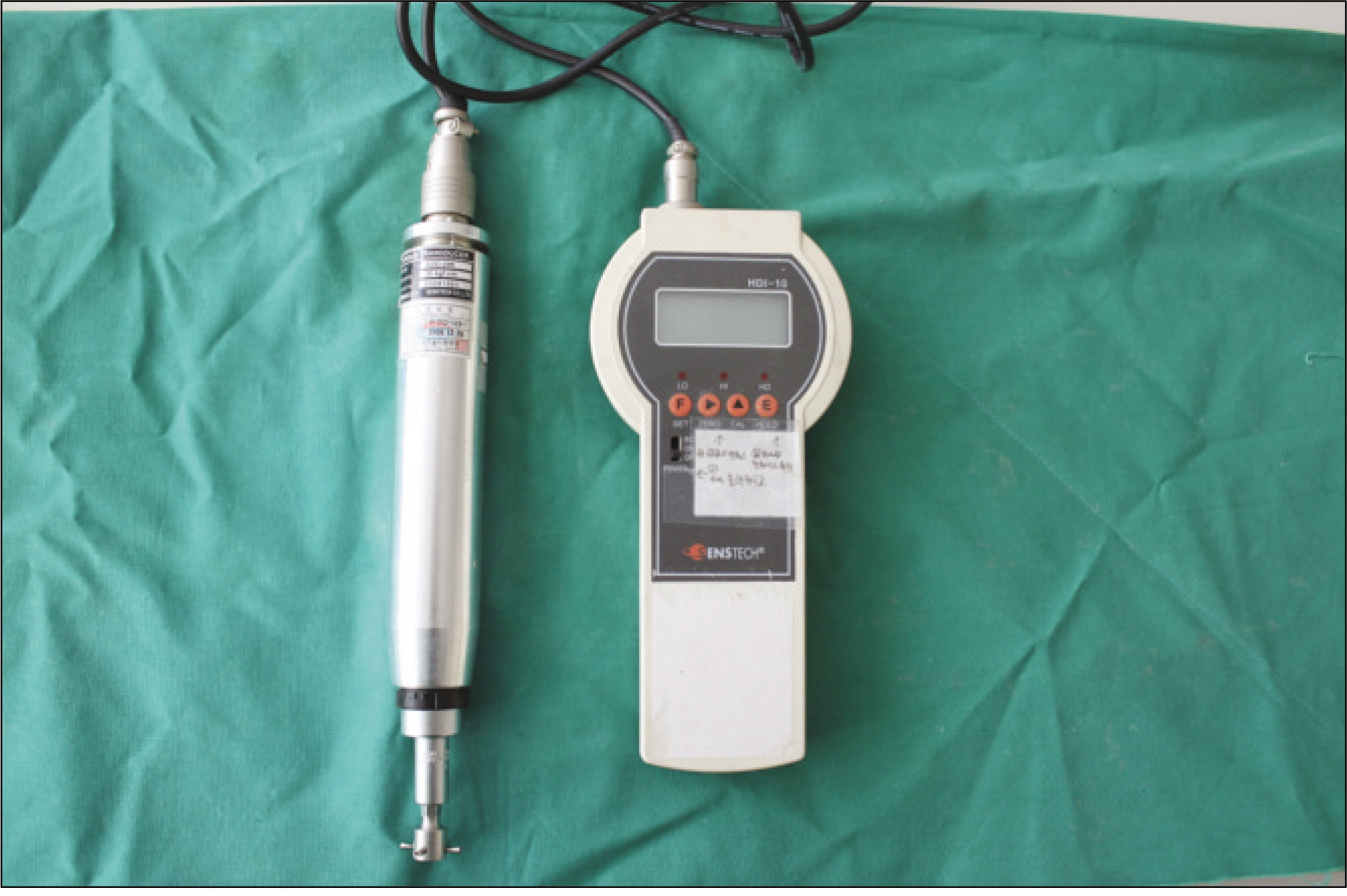
Fig. 1. Digital torque meter used to measure the peak value of resistance to reverse torque rotation in N cm.
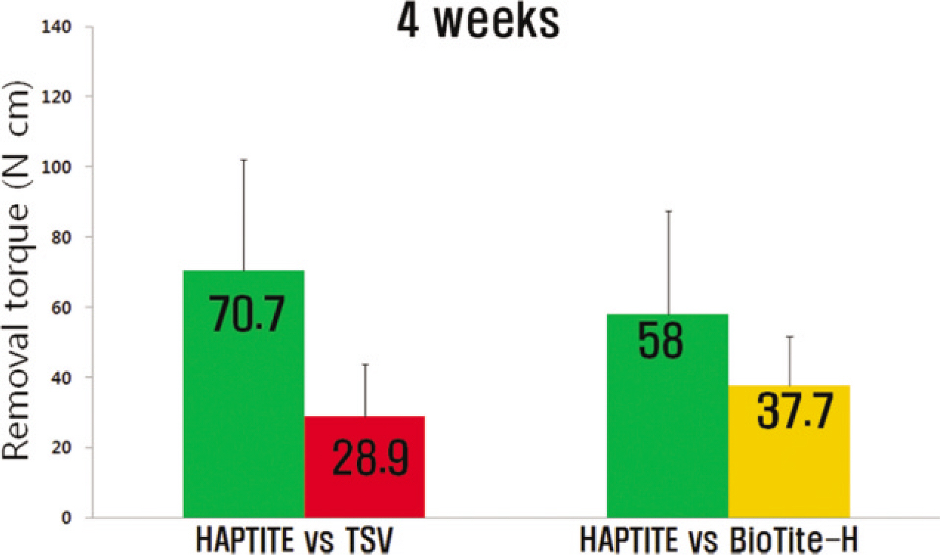
Fig. 2. Mean removal torque values (N cm) of implants 4 weeks after implantation.(TSV: Tapered Screw-Vent)

Fig. 3. Mean removal torque values (N cm) of implants 8 weeks after implantation.(TSV: Tapered Screw-Vent)
Cited by 1 articles
-
Assessment of the anterior loop of the inferior alveolar nerve via cone-beam computed tomography
Baratollah Shaban, Amin Khajavi, Nasim Khaki, Yones Mohiti, Tahere Mehri, Hamed Kermani
J Korean Assoc Oral Maxillofac Surg. 2017;43(6):395-400. doi: 10.5125/jkaoms.2017.43.6.395.
Reference
-
References
1. Bra�nemark PI, Adell R, Breine U, Hansson BO, Lindstro¨m J, Ohlsson A. Intraosseous anchorage of dental prostheses. I. Experimental studies. Scand J Plast Reconstr Surg. 1969; 3:81–100.2. Hutton JE, Heath MR, Chai JY, Harnett J, Jemt T, Johns RB, et al. Factors related to success and failure rates at 3-year follow-up in a multicenter study of overdentures supported by Bra�nemark implants. Int J Oral Maxillofac Implants. 1995; 10:33–42.3. Albrektsson T, Zarb G, Worthington P, Eriksson AR. The longterm efficacy of currently used dental implants: a review and proposed criteria of success. Int J Oral Maxillofac Implants. 1986; 1:11–25.4. Shirakura M, Fujii N, Ohnishi H, Taguchi Y, Ohshima H, Nomura S, et al. Tissue response to titanium implantation in the rat maxilla, with special reference to the effects of surface conditions on bone formation. Clin Oral Implants Res. 2003; 14:687–96.
Article5. Franchi M, Fini M, Martini D, Orsini E, Leonardi L, Ruggeri A, et al. Biological fixation of endosseous implants. Micron. 2005; 36:665–71.
Article6. Predecki P, Auslaender BA, Stephan JE, Mooney VL, Stanitski C. Attachment of bone to threaded implants by ingrowth and mechanical interlocking. J Biomed Mater Res. 1972; 6:401–12.7. Cook SD, Kay JF, Thomas KA, Jarcho M. Interface mechanics and histology of titanium and hydroxylapatite-coated titanium for dental implant applications. Int J Oral Maxillofac Implants. 1987; 2:15–22.8. Vercaigne S, Wolke JG, Naert I, Jansen JA. Histomorphometrical and mechanical evaluation of titanium plasma-spray-coated implants placed in the cortical bone of goats. J Biomed Mater Res. 1998; 41:41–8.
Article9. Buser D, Schenk RK, Steinemann S, Fiorellini JP, Fox CH, Stich H. Influence of surface characteristics on bone integration of titanium implants. A histomorphometric study in miniature pigs. J Biomed Mater Res. 1991; 25:889–902.
Article10. Cochran DL, Schenk RK, Lussi A, Higginbottom FL, Buser D. Bone response to unloaded and loaded titanium implants with a sandblasted and acid-etched surface: a histometric study in the canine mandible. J Biomed Mater Res. 1998; 40:1–11.
Article11. S � balle K, Hansen ES, Brockstedt-Rasmussen H, Pedersen CM, Bu¨nger C. Hydroxyapatite coating enhances fixation of porous coated implants. A comparison in dogs between press fit and noninterference fit. Acta Orthop Scand. 1990; 61:299–306.12. Block MS, Kent JN, Kay JF. Evaluation of hydroxylapatite-coated titanium dental implants in dogs. J Oral Maxillofac Surg. 1987; 45:601–7.
Article13. Wennerberg A, Albrektsson T, Andersson B, Krol JJ. A histomorphometric and removal torque study of screw-shaped titanium implants with three different surface topographies. Clin Oral Implants Res. 1995; 6:24–30.14. Kumar M, Dasarathy H, Riley C. Electrodeposition of brushite coatings and their transformation to hydroxyapatite in aqueous solutions. J Biomed Mater Res. 1999; 45:302–10.
Article15. Baker D, London RM, O'Neal R. Rate of pull-out strength gain of dual-etched titanium implants: a comparative study in rabbits. Int J Oral Maxillofac Implants. 1999; 14:722–8.16. Masuda T, Yliheikkila¨ PK, Felton DA, Cooper LF. Generalizations regarding the process and phenomenon of osseointegration. Part I. In vivo studies. Int J Oral Maxillofac Implants. 1998; 13:17–29.17. Cooper LF, Masuda T, Yliheikkila¨ PK, Felton DA. Generalizations regarding the process and phenomenon of osseointegration. Part II. In vitro studies. Int J Oral Maxillofac Implants. 1998; 13:163–74.18. Rich A, Harris AK. Anomalous preferences of cultured macrophages for hydrophobic and roughened substrata. J Cell Sci. 1981; 50:1–7.
Article19. Johansson C, Albrektsson T. Integration of screw implants in the rabbit: a 1-year follow-up of removal torque of titanium implants. Int J Oral Maxillofac Implants. 1987; 2:69–75.20. Sakakura CE, Margonar R, Holzhausen M, Nociti FH Jr, Alba RC Jr, Marcantonio E Jr. Influence of cyclosporin A therapy on bone healing around titanium implants: a histometric and biome-chanic study in rabbits. J Periodontol. 2003; 74:976–81.
Article21. Sennerby L, Thomsen P, Ericson LE. A morphometric and bio-mechanic comparison of titanium implants inserted in rabbit cortical and cancellous bone. Int J Oral Maxillofac Implants. 1992; 7:62–71.22. Ivanoff CJ, Widmark G, Johansson C, Wennerberg A. Histologic evaluation of bone response to oxidized and turned titanium micro-implants in human jawbone. Int J Oral Maxillofac Implants. 2003; 18:341–8.23. Roberts WE, Garreto LP. Bone physiology and metabolism. Misch CE, editor. Contemporary implant dentistry. 2nd ed.St. Louis: Mosby Co.;1999. p. 234–5.24. Misch CM. Hydroxylapatite-coated implants. Design considerations and clinical parameters. N Y State Dent J. 1993; 59:36–41. Erratum in: N Y State Dent J 1993;59: 12.25. Davies JE, Lowenberg B, Shiga A. The bone-titanium interface in vitro. J Biomed Mater Res. 1990; 24:1289–306.26. Yang Y, Bumgardner JD, Cavin R, Carnes DL, Ong JL. Osteoblast precursor cell attachment on heat-treated calcium phosphate coatings. J Dent Res. 2003; 82:449–53.
Article27. Cle`ries L, Ferna′ndez-Pradas JM, Sardin G, Morenza JL. Dissolution behaviour of calcium phosphate coatings obtained by laser ablation. Biomaterials. 1998; 19:1483–7.
Article
- Full Text Links
-
- Actions
-
Cited
- CITED
-
- Close
- Share
-
- Similar articles
-
- Comparing the precision of panoramic radiography and cone-beam computed tomography in avoiding anatomical structures critical to dental implant surgery: A retrospective study
- Implant Stability Change Measurement of Flaplessly Placed Immediate Implant on Mandibular First Molar
- Positioning errors of dental implants and their associations with adjacent structures and anatomical variations: A CBCT-based study
- Analysis and evaluation of relative positions of mandibular third molar and mandibular canal impacts
- Accuracy of digital surgical guides for dental implants
