A lateral approach to the maxillary sinus for simultaneous extraction of an ankylosed maxillary molar and sinus graft: a case report
- Affiliations
-
- 1Department of Advanced General Dentistry, College of Dentistry, Yonsei University, Seoul, Korea. wonse@yuhs.ac
- KMID: 2189707
- DOI: http://doi.org/10.5125/jkaoms.2012.38.2.110
Abstract
- Ankylosed tooth is defined as 'the discontinuance of normal passive tooth eruption without any mechanical barrier'. Ankylosed tooth treatment is a challenge to dental clinicians. In treatment of maxillary molar ankylosis cases there are risks of oro-antral fistula, displacement of root fragments into the maxillary sinus, as well as the necessity for providing additional sinus bone augmentation for future implant placement. In this study, we suggested a new technique using a piezoelectric device and a lateral side approach to the maxillary sinus leading to the simultaneous removal of the ankylosed maxillary molar and sinus grafting for the purpose of implant site development.
Keyword
MeSH Terms
Figure
-
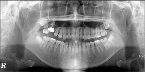
Fig. 1 Panoramic radiograph taken at the patient's first visit. An ankylosed right maxillary first molar is noted with full gold crown and root canal.

Fig. 2 Periapical view showing root resorption and loss of lamina dura around the infraoccluded tooth.

Fig. 3 Computed tomography evaluation showing the fusion of the tooth and alveolar bone segments; this fused apparatus is connected to the maxillary sinus septa.
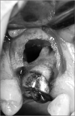
Fig. 4 Lateral approach to the ankylosed tooth after the removal of the anterior wall of the maxillary sinus. The Schneiderian (sinus) membrane was carefully detached from the root surface.
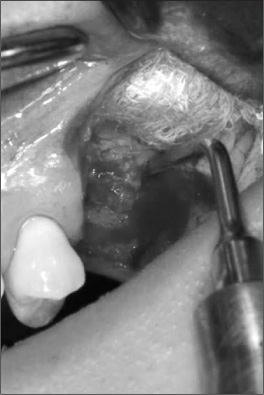
Fig. 5 A piezoelectric device was used to cut the residual fused tooth-bone segment to preserve sinus membrane integrity.
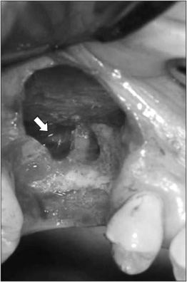
Fig. 6 The ankylosed tooth was completely removed. Some root-canal filling material was observed over the apex of the adjacent teeth (arrow).
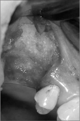
Fig. 7 Allogenic graft material was grafted at the defect.
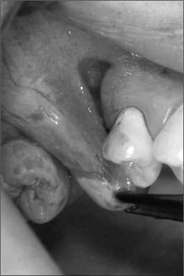
Fig. 8 A buccal advancement flap was formed with periosteal releasing incision for closure.
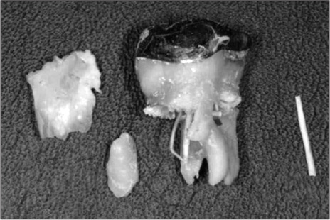
Fig. 9 The extracted tooth.
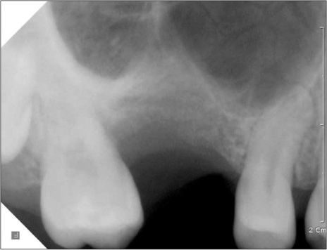
Fig. 10 The extracted tooth.
Reference
-
1. Biederman W. Etiology and treatment of tooth ankylosis. Am J Orthodont. 1962. 48:670–684.
Article2. Andersson L, Blomlöf L, Lindskog S, Feiglin B, Hammarström L. Tooth ankylosis. Clinical, radiographic and histological assessments. Int J Oral Surg. 1984. 13:423–431.3. Takahashi T, Takagi T, Moriyama K. Orthodontic treatment of a traumatically intruded tooth with ankylosis by traction after surgical luxation. Am J Orthod Dentofacial Orthop. 2005. 127:233–241.
Article4. Altuğ HA, Sahin S, Sencimen M, Dogan N. Extraction of upper first molar resulting in fracture of maxillary tuberosity. Dent Traumatol. 2009. 25:e1–e2.
Article5. Shultz RE, Theisen FC, Dunlap CL. Herniation of the antral membrane through an extraction site. Report of a case. Oral Surg Oral Med Oral Pathol. 1991. 71:280–282.6. Sethi A, Cariappa KM, Chitra A. Root fragment in the ostium of the maxillary sinus. Br J Oral Maxillofac Surg. 2009. 47:572–573.
Article7. González-García A, Diniz-Freitas M, Somoza-Martín M, García-García A. Ultrasonic osteotomy in oral surgery and implantology. Oral Surg Oral Med Oral Pathol Oral Radiol Endod. 2009. 108:360–367.
Article8. Landes CA, Stübinger S, Rieger J, Williger B, Ha TK, Sader R. Critical evaluation of piezoelectric osteotomy in orthognathic surgery: operative technique, blood loss, time requirement, nerve and vessel integrity. J Oral Maxillofac Surg. 2008. 66:657–674.
Article9. Stübinger S, Kuttenberger J, Filippi A, Sader R, Zeilhofer HF. Intraoral piezosurgery: preliminary results of a new technique. J Oral Maxillofac Surg. 2005. 63:1283–1287.
Article10. Torrella F, Pitarch J, Cabanes G, Anitua E. Ultrasonic ostectomy for the surgical approach of the maxillary sinus: a technical note. Int J Oral Maxillofac Implants. 1998. 13:697–700.11. Vercellotti T. Piezoelectric surgery in implantology: a case report--a new piezoelectric ridge expansion technique. Int J Periodontics Restorative Dent. 2000. 20:358–365.12. Vercellotti T, De Paoli S, Nevins M. The piezoelectric bony window osteotomy and sinus membrane elevation: introduction of a new technique for simplification of the sinus augmentation procedure. Int J Periodontics Restorative Dent. 2001. 21:561–567.13. Walmsley AD, Walsh TF, Laird WR, Williams AR. Effects of cavitational activity on the root surface of teeth during ultrasonic scaling. J Clin Periodontol. 1990. 17:306–312.
Article14. Rosano G, Taschieri S, Gaudy JF, Del Fabbro M. Maxillary sinus vascularization: a cadaveric study. J Craniofac Surg. 2009. 20:940–943.15. Mardinger O, Abba M, Hirshberg A, Schwartz-Arad D. Prevalence, diameter and course of the maxillary intraosseous vascular canal with relation to sinus augmentation procedure: a radiographic study. Int J Oral Maxillofac Surg. 2007. 36:735–738.
Article16. Abrahams JJ, Hayt MW, Rock R. Sinus lift procedure of the maxilla in patients with inadequate bone for dental implants: radiographic appearance. AJR Am J Roentgenol. 2000. 174:1289–1292.
Article17. Sánchez-Recio C, Peñarrocha-Diago M, Peñarrocha-Diago M, Peñarrocha-Oltra D. Maxillary sinus lift performed using ultrasound. Evaluation of 21 patients. Med Oral Patol Oral Cir Bucal. 2010. 15:e371–e374.18. Smiler DG, Johnson PW, Lozada JL, Misch C, Rosenlicht JL, Tatum OH Jr, et al. Sinus lift grafts and endosseous implants. Treatment of the atrophic posterior maxilla. Dent Clin North Am. 1992. 36:151–186.19. Chaushu S, Shapira J, Heling I, Becker A. Emergency orthodontic treatment after the traumatic intrusive luxation of maxillary incisors. Am J Orthod Dentofacial Orthop. 2004. 126:162–172.
Article20. Raghoebar GM, Boering G, Booy K, Vissink A. Treatment of the retained permanent molar. J Oral Maxillofac Surg. 1990. 48:1033–1038.
Article
- Full Text Links
-
- Actions
-
Cited
- CITED
-
- Close
- Share
-
- Similar articles
-
- Maxillary sinus pneumatization after maxillary molar extraction assessed with cone beam computed tomography
- Combined Sinus Floor and Alveolar Ridge Augmentation Simultaneously Performed with Extraction of Ankylosed Maxillary Molar: A Case Report
- Delayed Occurrence of Maxillary Sinusitis after Simultaneous Maxillary Sinus Augmentation and Implant: A Case Report and Literature Review
- Positional relationship between the maxillary sinus floor and the apex of the maxillary first molar using cone beam computed tomograph
- Implant placement simultaneously sinus augmentation using crestal approach in severely atrophic maxilla; minimally invasive approach
