J Korean Orthop Assoc.
2007 Oct;42(5):644-652. 10.4055/jkoa.2007.42.5.644.
Study of Bone Quality and Growth Characteristics of Growth Plate following Limb Transplantation between Animals of Different Ages-results of an Experimental Study on Male Syngeneic Rats
- Affiliations
-
- 1Department of Orthopedic Surgery, College of Medicine, Korea University Guro Hospital, Korea. spine@korea.ac.kr
- 2Department of Internal Medicine, College of Medicine, Ewha Womans University, Seoul, Korea.
- KMID: 2186472
- DOI: http://doi.org/10.4055/jkoa.2007.42.5.644
Abstract
-
BACKGROUND: Osteoporosis of transplanted/grafted bone is well known in the immediate post bone transplantation/bone grafting period. In limb transplantation, the growth plates in the transplanted limbs retain their longitudinal growth properties. However, there is a paucity of reports on what happens to the bone and the growth potential of the growth plate when limb transplantation between a juvenile donor and an adult recipient is performed.
MATERIALS AND METHODS
Ten juvenile to juvenile hind limb transplants and five juvenile to adult hind limb transplants were performed in male syngeneic Lewis rats. Osteoporosis in the isochronograft as well as the heterochronograft limbs was measured by 3D micro-CT. In addition, the increase in tibial length, after transplantation was measured and compared with the increase in the tibial length of the opposite non-operated limbs.
RESULTS
The 3D CT parameters indicate a significantly inferior bone quality in the heterochronografts compared with the isochronografts. After transplantation, the increase in the tibial length of the isochronografts was similar the increase in length of the opposite juvenile non operated tibiae and the heterochronograft tibias.
CONCLUSION
Age is a significant factor that affects the bone quality, resulting in post transplant osteoporosis in heterochronografts compared with isochronografts. However, the growth plate after transplantation remains unaffected by the difference in age and continues to grow at its own inherent rate in adult recipients as it does in the juvenile recipients.
Keyword
MeSH Terms
Figure
-

Fig. 1 Radiogram of the rat hindlimbs taken 3 weeks after transplantation-tibial lengths of both the transplanted and opposite non operated limb were measured.
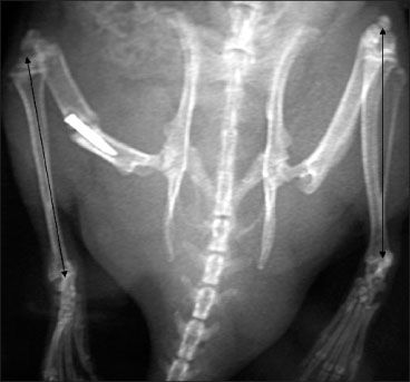
Fig. 2 Radiograph of rat hindlimbs 10 weeks after transplantation showing an increase in the length of the tibias of the transplanted limbs as well as the opposite non operated limbs.
Reference
-
1. Burchardt H. The biology of bone graft repair. Clin Orthop Relat Res. 1983. 174:28–42.
Article2. Burchardt H, Enneking WF. Transplantion of bone. Surg Clin North Am. 1978. 58:403–427.3. Kline SC, Hotchkiss RN, Randolph MA, Weiland AJ. Study of growth kinetics and morphology in limbs transplanted between animals of different ages. Plast Reconstr Surg. 1990. 85:273–280.
Article4. Steinberg EL, Luger E, Zwas T, Katznelson A. Very long-term radiographic and bone scan results of frozen autograft and allograft bone grafting in 17 patients (20 grafts) a 30- to 35-year follow-up. Cell Tissue Bank. 2004. 5:97–104.
Article5. Green H, Morikawa M, Nixon T. A dual effector theory of growth-hormone action. Differentiation. 1985. 29:195–198.
Article6. Kan KW, Cruess RL, Posner BI, Guyda HJ, Solomon S. Hormone receptors in the epiphysial cartilage. J Endocrinol. 1984. 103:125–131.
Article7. Nilsson A, Isgaard J, Lindahl A, Dahlstrom A, Skttner A, Isaksson OG. Regulation by growth hormone of number of chondrocytes containing IGF-I in rat growth plate. Science. 1986. 233:571–574.
Article8. Ogden JA, Southwick WO. Endocrine dysfunction and slipped capital femoral epiphysis. Yale J Biol Med. 1977. 50:1–16.9. Trippel SB, Van Wyk JJ, Mankin HJ. Localization of somatomedin-C binding to bovine growth-plate chondrocytes in situ. J Bone Joint Surg Am. 1986. 68:897–903.
Article10. Chiu HY, Harii K. Morphologic and growth alterations of epiphyseal plate after isohistogeneic transfer of young rat limb to adult rat. J Reconstr Microsurg. 1988. 4:103–111.
Article11. Drzewiecki AE, Randolph MA, Hotchkiss RH, Weiland AJ. Vascularized growth-plate transplantation: a comparative study in the rat. J Reconstr Microsurg. 1992. 8:93–100.
Article12. Innocenti M, Delcroix L, Manfrini M, Ceruso M, Capanna R. Vascularized proximal fibular transfer for distal radial reconstruction. J Bone Joint Surg Am. 2005. 87:Suppl 1. (Pt 2):237–246.13. Innocenti M, Delcroix L, Romano GF. Epiphyseal transplant: harvesting technique of the proximal fibula based on the anterior tibial artery. Microsurgery. 2005. 25:284–292.
Article14. Innocenti M, Delcroix L, Manfrini M, Ceruso M, Capanna R. Vascularized proximal fibular epiphyseal transfer for distal radial reconstruction. J Bone Joint Surg Am. 2004. 86:1504–1511.
Article15. Innocenti M, Ceruso M, Manfrini M, et al. Free vascularized growth-plate transfer after bone tumor resection in children. J Reconstr Microsurg. 1998. 14:137–143.
Article16. Vilkki SK. Distraction and micro vascular epiphysis transfer for radial club hand. J Hand Surg. 1995. 31:1.17. Donaldson HH. The rat: data and reference tables. 1992. 2nd ed. Philadelphia:18. Stevens DG, Boyer MI, Bowen CV. Transplantation of epiphyseal plate allografts between animals of different ages. J Pediatr Orthop. 1999. 19:398–403.
Article19. Glickman AM, Yang JP, Stevens DG, Bowen CV. Epiphyseal plate transplantation between sites of different growth potential. J Pediatr Orthop. 2000. 20:289–295.
Article
- Full Text Links
-
- Actions
-
Cited
- CITED
-
- Close
- Share
-
- Similar articles
-
- Vascularized Fibular Epiphysis and Epiphyseal Plate Transplantation
- An experimental study on the mandibular growth by the partial glossectomy of the rats
- Altered Cellular Kinetics in Growth Plate according to Alterations in Weight Bearing
- Bone Growth after Free Vascularized Grafting of the Upper Radius including its Epiphysis in Puppies
- Cultured Chondrocyte Transplantation in the Damaged Growth Plate
