The Usefulness of Calcium Sulfate in Treatment of Benign Bone Tumor
- Affiliations
-
- 1Department of Orthopeadic Surgery, Chonnam National University Medical School, Gwangju, Korea. rhamses@chol.com
- KMID: 2186469
- DOI: http://doi.org/10.4055/jkoa.2007.42.5.623
Abstract
-
PURPOSE: This study evaluated the results of the curettage and grafting of calcium sulfate for the treatment of a benign bone tumor to determine its efficacy as a bone graft substitute.
MATERIALS AND METHODS
Thirty six cases of calcium sulfate(Osteoset(R)) grafting for bone defect after curettage of benign bone tumor were evaluated. There were 21 males and 15 females with a mean age of 23 years (6-64). There were 23, 10 and 3 cases grafted with the Osteoset only, the Osteoset with allografts and the Osteoset with autografts, respectively. The average follow up duration was 19 months (12-49). The process of bone formation was observed and the times for graft absorption and complete bone formation were assessed. In addition, the time of bone formation was compared according to the patients' age, size of lesion, and grafting method, and the development of complications was observed.
RESULTS
Complete bone formation was observed in 34 (94.4%) out of 36 cases. The groups of younger patients, smaller size of lesion and grafting of Osteoset(R) only showed more rapid bone formation. However, there was no statistical significance. There was one case of soft tissue calcification observed.
CONCLUSION
Calcium sulfate is an effective substitute for an autogenous bone graft in the case of the treatment of a benign bone tumor, particularly in the case of an insufficient graft, such as in children and in those with a large bone defect. This method is a safe method that prevents complications in the donor site.
Keyword
MeSH Terms
Figure
-

Fig. 1 Pathologic fracture of the proximal femur associated with fibrous dysplasia in a 64-year-old female. (A) Preoperative plain radiograph shows a fracture of the proximal femur and radiolucent bone lesion at the fracture site. (B) Postoperative plain radiograph taken after plate fixation of the fracture and curettage and grafting of Osteoset® mixed with autograft. (C) Plain radiograph taken at 8 months after surgery shows bone union and complete healing of the previous bone lesion.
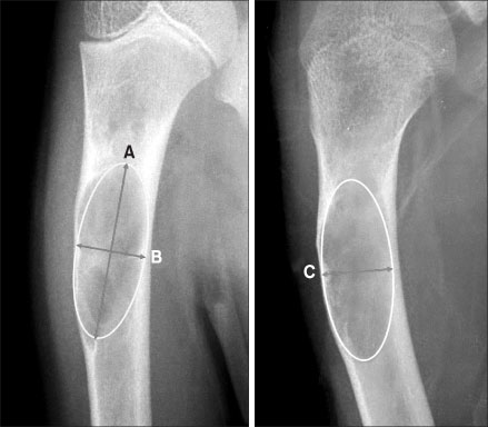
Fig. 2 Method for approximating the bone defect volume. Volume≒A×B×C.
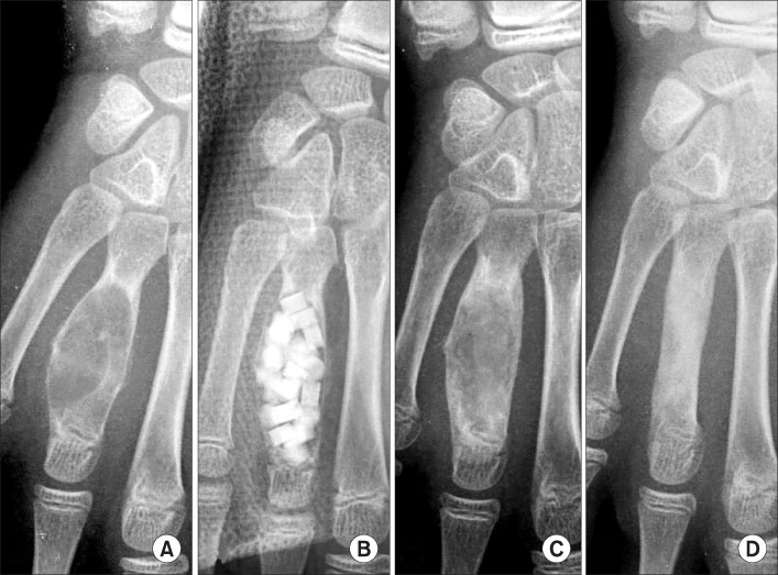
Fig. 3 Aneurysmal bone cyst of the metacarpal bone in a 13-year-old boy. (A) Preoperative plain radiograph shows an expansile cystic lesion of the fourth metacarpal bone. (B) Postoperative radiograph taken after curettage and grafting of Osteoset®. (C) Plain radiograph taken at 4 weeks after surgery shows the complete resorption of Osteoset®. (D) Plain radiograph taken at 5 months after surgery shows complete bone healing.

Fig. 4 Simple bone cyst of proximal humerus in a 10-year-old girl. (A) Preoperative plain radiograph shows radiolucent cystic lesion in the proximal humerus. (B) Postoperative radiograph taken after curettage and grafting of Osteoset®. (C) At 6 weeks after surgery, calcium sulfate was almost resorbed. (D) Plain radiograph taken at 18 months after surgery show a remnant lesion at a more distal portion. (E) A second operation was performed with curettage of remnant lesions and the grafting of Osteoset®. (F) Plain radiograph taken at 6 months after the second operation shows complete bone healing without a remnant lesion.

Fig. 5 Soft tissue calcification after the grafting of calcium sulfate. (A) Plain radiograph taken after 1 month after grafting the Osteoset® shows soft tissue calcification around the lesion. (B) The consolidation of calcification is seen at the 16 months follow up.
Reference
-
1. Dormans JP, Sankar WN, Moroz L, Erol B. Percutaneous intramedullay decompression, curettage, and grafting with medical-grade calcium sulfate pellets for unicameral bone cysts in children: a new minimally invasive technique. J Pediatr Orthop. 2005. 25:804–811.2. Dreesman H. Ueber Knochenplombierung. Biertr Klin Chir. 1892. 9:804–810.3. Fowler BL, Dall BE, Rowe DE. Complications associated with harvesting autogenous iliac bone graft. Am J Orthop. 1995. 24:895–903.4. Gaasbeek RD, Rijnberg WJ, van Loon CJ, Meyers H, Feith R. No local recurrence of enchondroma after curettage and plaster filling. Arch Orthop Trauma Surg. 2005. 125:42–45.
Article5. Gazdag AR, Lane JM, Glaser D, Forster RA. Alternatives to autogenous bone graft: efficacy and indications. J Am Acad Orthop Surg. 1995. 3:1–8.
Article6. Gitelis S, Haggard W, Piasecki P, Charters J, Turner T, Urban R. Use of a calcium sulfate-based bone graft substitute for benign bone lesions. Orthopedics. 2001. 24:162–166.
Article7. Han JS, Yoon KH, Ha JH. The use of calcium sulfate as a treatment of benign bone tumor. J Korean Bone Joint Tumor Soc. 2003. 9:31–37.8. Kelly CM, Wilkins RM, Gitelis S, Hartjen C, Watson JT, Kim PT. The use of a surgical grade calcium sulfate as a bone graft substitute: results of a multicenter trial. Clin Orthop Relat Res. 2001. 382:42–50.9. Mirzayan R, Panossian V, Avedian R, Forrester DM, Menendez LR. The use of calcium sulfate in the treatment of benign bone lesions. A preliminary report. J Bone Joint Surg Am. 2001. 83:355–358.10. Peltier LF. The use of plaster of Paris to fill defects in bone. Clin Orthop Relat Res. 1961. 21:1–31.11. Peltier LF, Jones RH. Treatment of unicameral bone cysts by curettage and packing with plaster-of-Paris Pellets. J Bone Joint Surg Am. 1978. 60:820–822.
Article12. Peters CL, Hines JL, Bachus KN, Craig MA, Bloebaum RD. Biological effects of calcium sulfate as a bone graft substitute in bovine metaphyseal defects. J Biomed Mater Res A. 2006. 76:456–462.13. Ricci JL, Rosenblum SF, Brezenoff L, Blumnethal NC. Stimulation of bone ingrowth into an implantable chamber through the use of rapidly resorbing calcium sulfate hemihydrate. 1992. In : Transactions of the Fourth World Biomaterials Congress; April; Berlin. European Society for Biomaterials;24–28.14. Rougraff BT. Bone graft alternatives in the treatment of benign bone tumors. Instr Course Lect. 2005. 54:505–512.15. Sidqui M, Collin P, Vitte C, Forest N. Osteoblast adherence and resorption activity of isolated osteoclasts on calcium sulphate hemihydrate. Biomaterials. 1995. 16:1327–1332.
Article16. Turner TM, Urban RM, Gitelis S, et al. Efficacy of calcium sulfate, a synthetic bone graft material, in healing a large canine medullary defect. Trans Ortho Res Soc. 1999. 45:522.
- Full Text Links
-
- Actions
-
Cited
- CITED
-
- Close
- Share
-
- Similar articles
-
- The Use of Calcium Sulfate as a Bone Substitute
- An experimental study on the effect of calcium sulfate on bone regeneration
- Tumor Necrosis Factor- and Resorption of Calcium Sulfate Used as a Bone Graft Substitute in Spinal Fusion in Rabbits
- Effect of composite of bone morphogenetic protein and plaster of paris on healing of bone defect in the rat tibia
- Efficacy of Calcium Sulfate Pellets as Bone Graft Substitute in Lumbar Posterolateral Fusion
