J Korean Orthop Assoc.
2009 Oct;44(5):507-513. 10.4055/jkoa.2009.44.5.507.
Effects of the Amount of Proximal Tibia Resection on the Bone Strength of Prepared Bone Surface: A FEM Study
- Affiliations
-
- 1Department of Orthopedic Surgery, Gongju Mediccal Center, Gongju, Korea. oskkk7@yahoo.co.kr
- 2Department of Orthopedic Surgery, Ajou University College of Medicine, Suwon, Korea.
- KMID: 2186309
- DOI: http://doi.org/10.4055/jkoa.2009.44.5.507
Abstract
- PURPOSE
We wanted to evaluate the mechanical strength of proximal tibia as resection distance increased from the joint surface. MATERIALS AND METHODS: We obtained the CT images of twenty knee osteoarthritis patients undergoing total knee arthroplasty. The finite element models were created based on the computed tomography images. The 8-node hexahedron element was made from BIONIX(TM) (CANTIBio. Co, Suwon, Korea), which is automatic mesh generation software program. The finite element model of the proximal tibia was resected at 6 mm, 8 mm, 10 mm, 12 mm, 15 mm and 18 mm from the lateral joint surface. A 1% strain rate was applied to a model by using HyperMesh(TM) software (Altair Engineering. Inc, Seattle, USA). The ultimate stress was calculated from the finite element analysis with using ANSYS 9.0 (ANSYS. Inc, Orlando, USA). RESULTS: The mean ultimate stress was 906.84 MPa, 877.22 MPa, 895.93 Mpa, 852.70 MPa, 742.90 Mpa and 585.51 Mpa at the 6 mm, 8 mm, 10 mm, 12 mm, 15 mm and 18 mm resection levels. As compare to the 6 mm resection level, the bone strengths at 15 mm and 18 mm were decreased with statistical significance (15 mm: p=0.005, 18 mm: p=0.000). CONCLUSION: The ultimate stress was decreased as the resection distance increased from the joint surface. But within a 12 mm resection distance from the lateral condyle articular surface of the tibia, the ultimate stress was not significantly decreased (p>0.05).
MeSH Terms
Figure
-

Fig. 1 Segmentation method by Bionix program on proximal tibia.
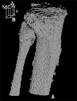
Fig. 2 Volumetric mesh model from computed tomographic data. (A) Results of region-growing algorithm. (B) Voxel size: X: 0.163 mm, Y: 0.163 mm, Z: 1.000 mm.
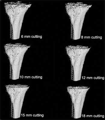
Fig. 3 The hexahedron mesh models of proximal tibia at each resection.

Fig. 4 Boundary condition. A strain was applied at the top face (◯) while displacement was constrained at the bottom face (◯).
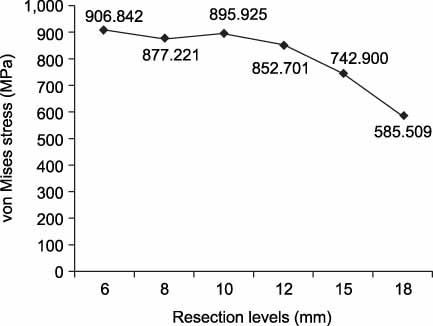
Fig. 5 Mean value of ultimate stress at each resection level.
Reference
-
1. Mckinley TO, Bay BK. Trabecular bone strain changes associated with subchondral stiffening of the proximal tibia. J Biomech. 2003. 36:155–163.
Article2. Chaput CD, Weeden SH, Hyman WA, Hitt KD. Mechanical bone strength of the tibial resection surface at increasing distance from the joint line in total knee arthroplasty. J Surg Orthop Adv. 2004. 13:195–198.3. Goldstein SA, Wilson DL, Sonstegard DA, Matthews LS. The mechanical properties of human tibial trabecular bone as a function of metaphyseal location. J Biomech. 1983. 16:965–969.
Article4. Harada Y, Wevers HW, Cooke TD. Distribution of bone strength in the proximal tibia. J Arthroplasty. 1988. 3:167–175.
Article5. Hvid I. Mechanical strength of trabecular bone at the knee. Dan Med Bull. 1988. 35:345–365.6. Hvid I, Jensen J, Nielsen S. Bone strength measurements at the proximal tibia. Penetration tests and epiphyseal compressive strength. Int Orthop. 1986. 10:271–275.7. van Rietbergen B, Weinans H, Huiskes R, Polman BJW. Computational strategies for iterative solutions of large FEM applications employing voxel data. Int J Num Meth Eng. 1998. 39:2743–2767.
Article8. Odgaard A, Linde F. The underestimation of Young's modulus in compressive testing of cancellous bone specimens. J Biomech. 1991. 24:691–698.
Article9. van Rietbergen B, Ulrich D, Pistoia W, Huiskes R, Ruegsegger P. Trabecular bone ultimate stress can be predicted from large-scale FE-analyses. J Biomech. 1998. 31:Suppl 1. 151.
Article10. van Rietbergen B, Weinans H, Huiskes R, Odgaard A. A new method to determine trabecular bone elastic properties and loading using micromechanical finite-element models. J Biomech. 1995. 28:69–81.
Article11. Ritter MA, Montgomery TJ, Zhou H, Keating ME, Faris PM, Meding JB. The clinical significance of proximal tibial resection level in total knee arthroplasty. Clin Orthop Relat Res. 1999. 360:174–181.
Article12. Bentzen SM, Hvid I, Jørgensen J. Mechanical strength of tibial trabecular bone evaluated by X-ray computed tomography. J Biomech. 1987. 20:743–752.
Article13. Bourne BC, van der Meulen MC. Finite element models predict cancellous apparent modulus when tissue modulus is scaled from specimen CT-attenuation. J Biomech. 2004. 37:613–621.
Article14. Sarathi Kopparti P, Lewis G. Influence of three variables on the stresses in a three-dimensional model of a proximal tibia-total knee implant construct. Biomed Mater Eng. 2007. 17:19–28.15. Majumdar S, Kothari M, Augat P, et al. High-resolution magnetic resonance imaging: Three-dimensional trabecular bone architecture and biomechanical properties. Bone. 1998. 22:445–454.
Article
- Full Text Links
-
- Actions
-
Cited
- CITED
-
- Close
- Share
-
- Similar articles
-
- Acute Shortening and Gradual Lengthening for a Comminuted Tibia Fracture with Massive Bone and Soft Tissue Defect: Case Report
- Radiological , Biomechanical and Histological Analysis on the Surgical Treatment of Bone Defect in Rabbit Tibia using Glass Ceramics
- A clinical comparative study between anterior iliac and proximal tibial metaphysis particulated cancellous bone grafts
- The Results of Hemicortical Resection for Malignant Bone Tumor
- The Bone Mineral Density of the Proximal Tibia, Lumbar Spine and Proximal Femur and Its Correlation with the Alignment of the Lower Extremity in Knee Osteoarthritic Patients
