J Korean Orthop Assoc.
2010 Feb;45(1):24-30. 10.4055/jkoa.2010.45.1.24.
First Metatarsal Dorsal Close Wedge Osteotomy Combined with Medial Cuneiform Plantar Open Wedge Osteotomy for the Treatment of a Cavus Foot
- Affiliations
-
- 1Department of Orthopaedic Surgery, Chonbuk National University Medical School, Research Institute of Clinical Science, JeonJu, Korea. jrkeem@chonbuk.ac.kr
- KMID: 2185590
- DOI: http://doi.org/10.4055/jkoa.2010.45.1.24
Abstract
- PURPOSE
We wanted to analyze the results of the 1st metatarsal dorsal close wedge osteotomy (MTDW) combined with medical cuneiform plantar open wedge (MCPOW) for treating forefoot deformity of a cavus foot.
MATERIALS AND METHODS
We retrospectively analyzed 30 patients. Their mean age was 21.5 years (SD 10.6 years) and the average follow-up period was 2.3 years. Thirty-four cases of thirty patients were classified as group A, as classified by the 1st MTDW combined with the MCPOW, 16 feet (14 patients) were group B by the 1st MTDW or MCPOW, 12 feet (10 patients), and group C by triple arthrodesis, 6 feet (6 patients). We evaluated the ankle dorsiflexion, plantarflexion, heel alignment, and the Maryland foot score (MFS) preoperatively and the last follow-up, and we analyzed the radiologic Hibb, Meary, calcaneal pitch and tibiotalar angles.
RESULTS
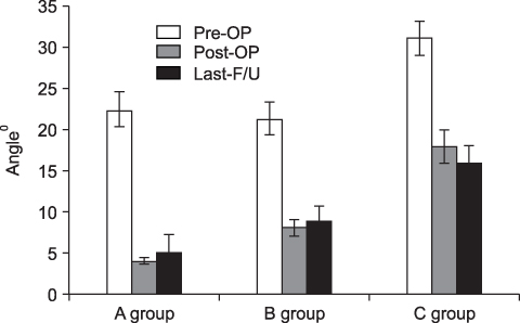
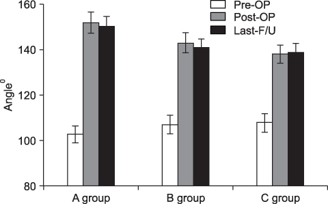
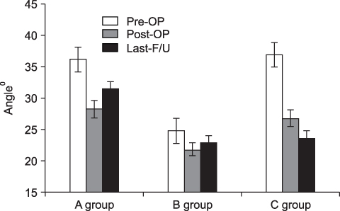
The ankle dorsiflexion (p=0.01), plantar flexion (p=0.03) and heel alignment (p=0.02) of group A were significantly improved more than that of groups B and C. The MFS of group A revealed better than group B and C (p=0.01). The Meary (p=0.01), Hibb (p=0.02) and calcaneal pitch angle (p=0.02) of group A were significantly improved more than that of groups B and C.
CONCLUSION
1st MTDW combined with MCPOW osteotomy that focuses at the apex of the deformity for correction of a cavus foot can obtain better clinical and radiological results than other surgical procedures.
Keyword
MeSH Terms
Figure
Reference
-
1. Mosca VS. The cavus foot. J Pediatr Ortho. 2001. 21:423–424.
Article2. Paulos L, Coleman SS, Samuelson KM. Pes cavovarus. Review of a surgical approach using selective soft-tissue procedures. J Bone Joint Surg Am. 1980. 62:942–953.
Article3. Sabir M, Lyttle D. Pathogenesis of pes cavus in Charcot-Marie-Tooth disease. Clin Orthop Relat Res. 1983. 175:173–178.
Article4. Alexander IJ, Johnson KA. Assessment and management of pes cavus in Charcot-Marie-tooth disease. Clin Orthop Relat Res. 1989. 246:273–281.
Article5. Azmaipairashvili Z, Riddle EC, Scavina M, Kumar SJ. Correction of cavovarus foot deformity in Charcot-Marie-Tooth disease. J Pediatr Orthop. 2005. 25:360–365.
Article6. Sanders R, Fortin P, DiPasquale T, Walling A. Operative treatment in 120 displaced intra articular calcaneal fractures. Results using a prognostic computed tomography scan classification. Clin Orthop Relat Res. 1993. 290:87–95.7. Sherman FC, Westin GW. Plantar release in the correction of deformities of the foot in childhood. J Bone Joint Surg Am. 1981. 63:1382–1389.
Article8. Jahss MH. Tarsometatarsal truncated-wedge arthrodesis for pes cavus and equinovarus deformity of the fore part of the foot. J Bone Joint Surg Am. 1980. 62:713–722.
Article9. Wicart P, Seringe R. Plantar opening-wedge osteotomy of cuneiform bones combined with selective plantar release and dwyer osteotomy for pes cavovarus in children. J Pediatr Orthop. 2006. 26:100–108.
Article10. Medhat MA, Karantz H. Neuropathic ankle joint in Charcot-Marie-Tooth disease after triple arthrodesis of the foot. Ortho Rev. 1988. 17:873–880.11. Wukich DK, Bowen JR. A long-term study of triple arthrodesis for correction of pes cavovarus in Charcot-Marie-Tooth disease. J Pediatr Orthop. 1989. 9:433–437.
Article12. Mubarak SJ, Van Valin SE. Osteotomies of the foot for cavus deformities in children. J Pediatr Orthop. 2009. 29:294–299.
Article13. Coleman SS, Chestnut WJ. A Simple test for hindfoot flexibility in the cavovarus foot. Clin Orthop. 1977. 123:60–62.
Article14. Dwyer FC. Osteotomy of the calcaneum for pes cavus. J Bone Joint Surg Br. 1959. 41:80–86.
Article15. Jahss MH. Evaluation of the cavus foot for orthopedic treatment. Clin Orthop Relat Res. 1983. 181:52–63.
Article16. Schwend RM, Drennan JC. Cavus foot deformity in children. J Am Acad Orthop Surg. 2003. 11:201–211.
Article17. Giannini S, Ceccarelli F, Benedetti MG, Faldini C, Grandi G. Surgical treatment of adult idiopathic cavus foot with plantar fasciotomy, naviculocuneiform arthrodesis, and cuboid osteotomy. A review of thirty-nine cases. J Bone Joint Surg Am. 2002. 84:Suppl. 62–69.18. Mann DC, Hsu JD. Triple arthrodesis in the treatment of fixed cavovarus deformity in adolescent patients with Charcot-Marie-Tooth disease. Foot Ankle. 1992. 13:1–6.
Article19. Wetmore RS, Drennan JC. Long-term results of triple arthrodesis in Charcot-Marie-Tooth disease. J Bone Joint Surg Am. 1989. 71:417–422.
Article20. Gould N. Surgery in advanced Charcot-Marie-Tooth disease. Foot Ankle. 1984. 4:267–273.
Article21. Japas LM. Surgical treatment of pes cavus by tarsal V-osteo-tomy. Preliminary report. J Bone Joint Surg Am. 1968. 50:927–944.
- Full Text Links
- Actions
-
Cited
- CITED
-
- Close
- Share
- Similar articles
-
- The Results of the First Ray Forefoot Osteotomy Using Low Profile Wedge Plate without a Bone Grafting for Pes Planus Correction
- Dorsal Wedge Osteotomy Using Bioabsorbable Pins for the Treatment of Freiberg's Disease
- Dorsal Closing Wedge Osteotomy in Freiberg's Disease
- Weil Osteotomy for Freiberg's Disease
- Half-wedge osteotomy and reverse repositioning for dorsal malunion distal radius fracture: a preliminary report with a case series


