Intra-Articular Osteochondroma of the Patella
- Affiliations
-
- 1Department of Orthopedic Surgery, Daegu Fatima Hospital, Daegu, Korea. hwan1260@naver.com
- KMID: 2185312
- DOI: http://doi.org/10.4055/jkoa.2012.47.6.468
Abstract
- Osteochondroma is one of the most common benign tumors. However, intra-articular occurrence is rare, especially in the knee joint; this is a case of osteochondroma forming bony bridge with the patella. The authors experienced a case of intra-articular osteochondroma of the left patella observed in a 58-year-old female, so we reported it with a literature review.
Keyword
MeSH Terms
Figure
-
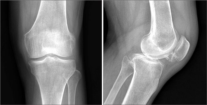
Figure 1 Preoperative plain radiographs of the left knee. Lateral view shows a mass-like lesion with bony shadow projecting from the inferior pole of patella.
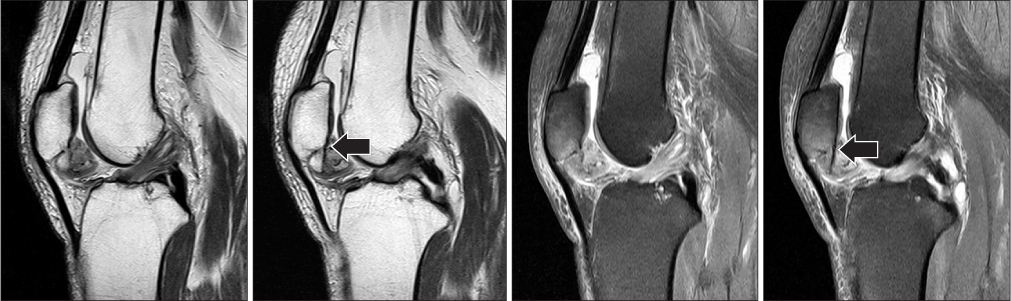
Figure 2 Magnetic resonance imaging of the lesion sagittal T1, T2-weighted images show cortical and cancellous bony connection between the inhomogenous mass with marginal cartilage shadow and the posteromedial corner of the inferior pole of patella directly (arrow).
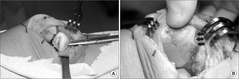
Figure 3 In operative field, the mass was rigidly connected with the inferior pole of patella through the bony bridge. There was no connection with infra-patellar fat pad, synovium or patellar tendon. The bony mass was excised clearly, (A) before and (B) after. The cut surface of bony bridge at the posteromedial corner (multi-arrow).
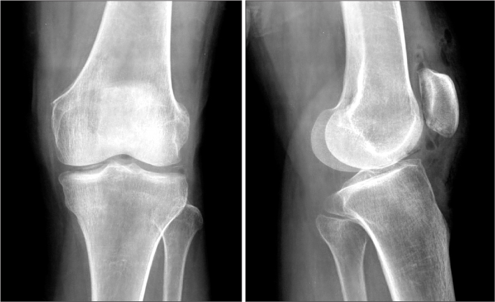
Figure 4 Postoperative plain radiographs show that intra-articular osteochondroma was clearly removed from the inferior pole of patella.
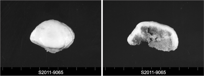
Figure 5 The excised specimen. This consists of ovoid polypoid bony tissue, measuring 3.2×2×2 cm. Cut section shows grayish thickened cartilage, of which cap thickness is 2 mm.
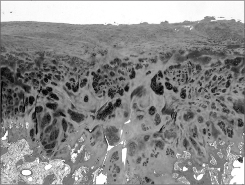
Figure 6 The microscopic finding of the excised mass (H&E, ×20). The typical findings of osteochondroma are seen.
Reference
-
1. Murphey MD, Choi JJ, Kransdorf MJ, Flemming DJ, Gannon FH. Imaging of osteochondroma: variants and complications with radiologic-pathologic correlation. Radiographics. 2000. 20:1407–1434.2. Reith JD, Bauer TW, Joyce MJ. Paraarticular osteochondroma of the knee: report of 2 cases and review of the literature. Clin Orthop Relat Res. 1997. (334):225–232.3. Jaffe HL. Tumors and tumorous conditions of the bones and joints. 1958. Philadelphia: Lea & Febiger;143–150. 558–567.4. Han CS, Jeong BO, So DH. Intra-articular osteochondroma of the knee. J Korean Bone Joint Tumor Soc. 2004. 10:147–151.5. Sohn SW, Kim IS. A case of intraarticular osteochondroma arising from patella. J Korean Orthop Assoc. 1998. 33:620–623.6. Gulati Y, Maheshwari A, Sharma V, Mattoo R, Arora D, Gupta N. Extraskeletal osteochondroma of the thigh: a case report. Acta Orthop Belg. 2005. 71:115–118.7. Lim Y, Kim ES, Shin JK, Kim BJ. Giant intraarticular osteochondroma in the knee joint of 14 years old athlete: report of 1 case. J Korean Knee Soc. 1993. 5:218–221.8. Moon MS, Woo YK, Yang SW. Intra-articular osteochondroma of the knee: a case report. J Korean Orthop Assoc. 1984. 19:735–737.9. Hagan PF, Schoenecker PL. Para-articular osteochondroma. Am J Orthop (Belle Mead NJ). 1995. 24:65–67.10. Lee KH, Kang SI, Park CS, Kim MK, Kim MS. Giant intra-articular osteochondroma of the knee: a case report. J Korean Orthop Assoc. 1990. 25:973–975.
- Full Text Links
-
- Actions
-
Cited
- CITED
-
- Close
- Share
-
- Similar articles
-
- Giant Intra-articular Osteochondroma of the Knee: A Case Report
- Intra - Articular Osteochondroma of the Knee: A Case Report
- A Case of Intraarticular Osteochondroma Arising from Patella
- Arthroscopic Excision of Intra-articular Osteochondroma of the Elbow: A Case Report
- Arthroscopic Excision of Solitary Intra-articular Osteochondroma of the Knee
