Factors Affecting Adjacent Level Ossification Development after Anterior Cervical Discectomy and Fusion
- Affiliations
-
- 1Department of Orthopedic Surgery, Presbyterian Medical Center, Jeonju, Korea. docby@hanmail.net
- KMID: 2185305
- DOI: http://doi.org/10.4055/jkoa.2012.47.6.425
Abstract
- PURPOSE
The purpose of this study is to analyze the affecting factors of adjacent level ossification development (ALOD) after anterior cervical discectomy and fusion.
MATERIALS AND METHODS
This study enrolled 75 patients who underwent anterior cervical discectomy and fusion and were followed-up for more than two years. Twenty-five patients were related with trauma and 47 patients were diagnosed as degenerative cervical disorder. We assessed the incidence, location and timing of ALOD, and compared the incidence of ossification between trauma and degenerative disease groups to know the effect of soft tissue damage. We also reviewed the correlation between the development of ossification at adjacent level and the factors, such as fusion level, age, operation time, duration of follow-up, and the presence of ossification of posterior longitudinal ligament (OPLL), as well as ossification of yellow ligament (OYL).
RESULTS
Ossification developed in 33 patients (44%). Five cases (15%) were detected during the first year after surgery, 10 (30%) cases detected during the second year after surgery, 13 (40%) between second and third year, and 5 (15%) cases of more than three years after surgery. Only the fusion level was related with the development of ossification statistically (p<0.001). Age, operation time, duration of follow-up, sex ratio, presence of OPLL, and OYL were not related with the incidence of ossification significantly. There was no significant difference in the incidence of ALOD between the trauma group and degenerative disease group (p=0.3625).
CONCLUSION
To detect ALOD, it needs a long time for follow-up after surgery. We thought that ALOD is affected by excessive loading at the adjacent level after fusion rather than severity of the soft tissue damage.
MeSH Terms
Figure
-

Figure 1 Radiographic parameters on lateral plane radiographs of the cervical spine. The plate-to-disc distances were measured from the tips of the plate to the (A) cephalad and (B) to the caudal, respectively.
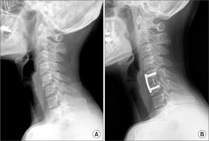
Figure 2 A 61-year-old female patient had anterior cervical discectomy and fusion with Polyethyletherketone (PEEK) cage (Stryker spine, USA) packed with autogenous iliac bone and plate stabilization for unilateral facet dislocation at C5-6. (A) Preoperative lateral radiograph. (B) Last follow-up radiograph (postoperative 57 months), it shows no change in adjacent levels.
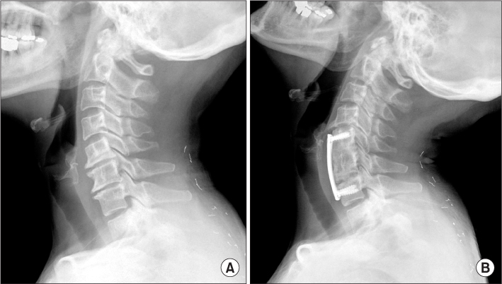
Figure 3 A 46-year-old female patient had anterior cervical discectomies and fusions with cages and plate for degenerative cervical spondylosis associated with disc herniation at C5-6, 6-7. (A) Preoperative lateral radiograph. (B) Last follow-up radiograph (postoperative 49 months), it shows ossification in cranial adjacent disc space.

Figure 4 A 40-year-old male patient had anterior cervical discectomies and fusions with cages and plate for degenerative cervical spondylosis associated with disc herniation at C5-6, 6-7. (A) Preoperative lateral radiograph. (B) After follow-up of 15 months it shows no definite change in adjacent level. (C) Lateral radiograph at 34 months after surgery shows ossification in caudal adjacent disc space.
Reference
-
1. Bohlman HH, Emery SE, Goodfellow DB, Jones PK. Robinson anterior cervical discectomy and arthrodesis for cervical radiculopathy. Long-term follow-up of one hundred and twenty-two patients. J Bone Joint Surg Am. 1993. 75:1298–1307.
Article2. Braunstein EM, Hunter LY, Bailey RW. Long term radiographic changes following anterior cervical fusion. Clin Radiol. 1980. 31:201–203.
Article3. Gore DR, Sepic SB. Anterior. Spine (Phila Pa 1976). 1984. 9:667–671.
Article4. Goto S, Mochizuki M, Kita T, et al. Anterior surgery in four consecutive technical phases for cervical spondylotic myelopathy. Spine (Phila Pa 1976). 1993. 18:1968–1973.
Article5. Goffin J, van Loon J, Van Calenbergh F, Plets C. Long-term results after anterior cervical fusion and osteosynthetic stabilization for fractures and/or dislocations of the cervical spine. J Spinal Disord. 1995. 8:500–508.
Article6. Kim KS. Long-term effects on the cervical spine after anterior locking plate fixation. J Korean Neurosurg Soc. 2001. 30:493–500.
Article7. Park JB, Cho YS, Riew KD. Development of adjacent-level ossification in patients with an anterior cervical plate. J Bone Joint Surg Am. 2005. 87:558–563.
Article8. Yang JY, Song HS, Lee M, Bohlman HH, Riew KD. Adjacent level ossification development after anterior cervical fusion without plate fixation. Spine (Phila Pa 1976). 2009. 34:30–33.9. Park JY, Zhang HY, Oh MC. New technical tip for anterior cervical plating: make hole first and choose the proper plate size later. J Korean Neurosurg Soc. 2011. 49:212–216.10. Smith GW, Robinson RA. The treatment of certain cervical-spine disorders by anterior removal of the intervertebral disc and interbody fusion. J Bone Joint Surg Am. 1958. 40:607–624.11. Park JB, Watthanaaphisit T, Riew KD. Timing of development of adjacent-level ossification after anterior cervical arthrodesis with plates. Spine J. 2007. 7:633–636.12. Modabber MR, Jupiter JB. Reconstruction for post-traumatic conditions of the elbow joint. J Bone Joint Surg Am. 1995. 77:1431–1446.
Article13. Garland DE, Orwin JF. Resection of heterotopic ossification in patients with spinal cord injuries. Clin Orthop Relat Res. 1989. (242):169–176.
Article14. Hasegawa M, Ohashi T, Uchida A. Heterotopic ossification around distal femur after total knee arthroplasty. Arch Orthop Trauma Surg. 2002. 122:274–278.15. Sperling JW, Cofield RH, Rowland CM. Heterotopic ossification after total shoulder arthroplasty. J Arthroplasty. 2000. 15:179–182.16. Purtill JJ, Eng K, Rothman RH, Hozack WJ. Heterotopic ossification. Incidence in cemented versus cementless total hip arthroplasty. J Arthroplasty. 1996. 11:58–63.
Article17. Mähring M. Segment changes in the cervical spine following cervical spondylodeses of unstable injuries. Unfallchirurgie. 1988. 14:247–258.
Article18. Nagata H, Schendel MJ, Transfeldt EE, Lewis JL. The effects of immobilization of long segments of the spine on the adjacent and distal facet force and lumbosacral motion. Spine (Phila Pa 1976). 1993. 18:2471–2479.
Article19. Döhler JR, Kahn MR, Hughes SP. Instability of the cervical spine after anterior interbody fusion. A study on its incidence and clinical significance in 21 patients. Arch Orthop Trauma Surg. 1985. 104:247–250.
Article20. Fuller DA, Kirkpatrick JS, Emery SE, Wilber RG, Davy DT. A kinematic study of the cervical spine before and after segmental arthrodesis. Spine (Phila Pa 1976). 1998. 23:1649–1656.
Article21. Garrido BJ, Wilhite J, Nakano M, et al. Adjacent-level cervical ossification after Bryan cervical disc arthroplasty compared with anterior cervical discectomy and fusion. J Bone Joint Surg Am. 2011. 93:1185–1189.
Article22. Aota Y, Kumano K, Hirabayashi S. Postfusion instability at the adjacent segments after rigid pedicle screw fixation for degenerative lumbar spinal disorders. J Spinal Disord. 1995. 8:464–473.23. Kolstad F, Nygaard ØP, Leivseth G. Segmental motion adjacent to anterior cervical arthrodesis: a prospective study. Spine (Phila Pa 1976). 2007. 32:512–517.
- Full Text Links
-
- Actions
-
Cited
- CITED
-
- Close
- Share
-
- Similar articles
-
- Risk Factors for “Adjacent-Level Ossification Development” Other Than Short Plate-to-Disc Distance and Clinical Implications for Adjacent-Segment Pathology
- Recycling of Cervical Artificial Disc for the Symptomatic Adjacent Segment Disorder Combined with Instability on Total Disc Replacement Area: A Case Report
- Degenerative Changes of Adjacent Segment Following Cervical Anterior fusion: Correlation with Preoperative MRI Findings of Adjacent Disc
- Analysis of Noninstrumented Anterior Cervical Discectomy and Interbody Fusion in Degenerative Cervical Disease
- A Comparison of Anterior Cervical Discectomy and Fusion versus Fusion Combined with Artificial Disc Replacement for Treating 3-Level Cervical Spondylotic Disease
