Analysis of Low-Energy Trochanter Fracture Using the Multiplanar Computed Tomography Image: Application for Intramedullary Nail Fixation
- Affiliations
-
- 1Department of Orthopaedic Surgery, Kosin University College of Medicine, Busan, Korea. jyujin2001@kosin.ac.kr
- 2Department of Orthopaedic Surgery, Gijang Hospital, Busan, Korea.
- KMID: 2184407
- DOI: http://doi.org/10.12671/jkfs.2015.28.3.155
Abstract
- PURPOSE
The purpose of this radiologic study was to evaluate the geographic patterns of low-energy trochanteric fractures using multiplanar computed tomography (CT) images for application of intramedullary nailing.
MATERIALS AND METHODS
In this study, 117 trochanteric fractures (stable fracture, 39 cases, unstable fractures, 78 cases) sustained from simple slip-down were assessed. The mean age was 78.4 years (range, 60-96 years). Multiplanar CT images were assessed for evaluation of geographic features of trochanteric fracture, and the fracture exit and geographic patterns were analyzed.
RESULTS
The medial and lateral exit of the trochanteric fracture showed no statistical difference by age, bone density, and comorbid disease. The exit was located at an average distance of 10.2 mm (range, 1.0-22.2 mm) from the tip of the greater trochanter (GT), and the medial exit, average distance of 8.1 mm (range, 0.0-18.3 mm) from the tip of the lesser trochanter. It was also found that there was no comminution around the anteromedial cortex of the fracture, and its contact loss was from fracture deformity.
CONCLUSION
Because of no comminution, the contact restoration of the anteromedial cortex resulted in correction of fracture deformity and reduction. Trochanteric nailing by GT tip could be fixed through the proximal fragment of the fracture because the lateral exit is placed at an average distance of 10.2 mm from the GT tip.
Figure
-
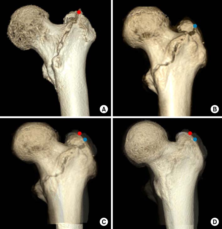
Fig. 1 Images of a trochanter fracture overlap each other (A, B) using Photoshop CS6® according to 50% opacity (C) and 20% opacity (D).

Fig. 2 Image of an overlapped trochanter fracture on a normal femur image. The lateral exit of the trochanteric fracture is located at an average distance of 10.2 mm without lateral wall fracture and not below the vastus ridge.

Fig. 3 Image of an overlapped trochanter fracture on a normal femur image. The medial exit is located at an average distance of 8.1 mm.
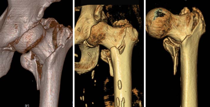
Fig. 4 The anteromedial cortex of the trochanter fracture was not comminuted, and however, showed several types.

Fig. 5 Immediate postoperative radiographs show that the proximal fragment of the trochanter fracture is displaced and widened inferiorly because of incorrect intramedullary nail insertion along the fracture site.
Reference
-
1. Kokoroghiannis C, Aktselis I, Deligeorgis A, Fragkomichalos E, Papadimas D, Pappadas I. Evolving concepts of stability and intramedullary fixation of intertrochanteric fractures--a review. Injury. 2012; 43:686–693.
Article2. Bridle SH, Patel AD, Bircher M, Calvert PT. Fixation of intertrochanteric fractures of the femur. A randomised prospective comparison of the gamma nail and the dynamic hip screw. J Bone Joint Surg Br. 1991; 73:330–334.
Article3. Haidukewych GJ. Intertrochanteric fractures: ten tips to improve results. J Bone Joint Surg Am. 2009; 91:712–719.4. Gotfried Y. Integrity of the lateral femoral wall in intertrochanteric hip fractures: an important predictor of a reoperation. J Bone Joint Surg Am. 2007; 89:2552–2553.
Article5. Im GI. Treatment of unstable intertrochanteric fractures. A comparison of proximal femoral nail and dynamic hip screw. J Korean Hip Soc. 2007; 19:36–44.
Article6. Shrout PE, Fleiss JL. Intraclass correlations: uses in assessing rater reliability. Psychol Bull. 1979; 86:420–428.
Article7. Jung GH. Treatment of the intertrochanteric femoral fracture with proximal femoral nail: nailing using the provisional K-wire fixation. J Korean Fract Soc. 2011; 24:223–229.
Article8. Oh JK, Hwang JH. Osteoporotic pertrochanteric fracture: IM nailing. J Korean Fract Soc. 2009; 22:56–65.
Article9. Hak DJ, Bilat C. Avoiding varus malreduction during cephalomedullary nailing of intertrochanteric hip fractures. Arch Orthop Trauma Surg. 2011; 131:709–710.
Article10. Ostrum RF, Marcantonio A, Marburger R. A critical analysis of the eccentric starting point for trochanteric intramedullary femoral nailing. J Orthop Trauma. 2005; 19:681–686.
Article11. Im GI, Shin YW, Song YJ. Potentially unstable intertrochanteric fractures. J Orthop Trauma. 2005; 19:5–9.
Article12. Kukla C, Heinz T, Gaebler C, Heinze G, Vécsei V. The standard Gamma nail: a critical analysis of 1,000 cases. J Trauma. 2001; 51:77–83.
Article13. Chang WS, Zuckerman JD, Kummer FJ, Frankel VH. Biomechanical evaluation of anatomic reduction versus medial displacement osteotomy in unstable intertrochanteric fractures. Clin Orthop Relat Res. 1987; (225):141–146.
Article14. Carr JB. The anterior and medial reduction of intertrochanteric fractures: a simple method to obtain a stable reduction. J Orthop Trauma. 2007; 21:485–489.
Article
- Full Text Links
-
- Actions
-
Cited
- CITED
-
- Close
- Share
-
- Similar articles
-
- Flexible Intramedullary Nail Fixation of Pediatric Femoral Shaft Fracture
- Retrieval of a Broken Intramedullary Nail after Refracture of Femoral Shaft: A case report
- Difficulty in Removal of a Femoral Intramedullary Nail: The Geometry of the Distal End of the Nail
- Femoral Neck Fracture After Removal of the Intramedullary Nail for the Fixation of an Intertrochanteric Fracture: Report of 2 Cases
- Acase of Fatigue Fracture of the Interlocking nail in the Treatment of Fracture of the Femoral Shaft
