J Dent Rehabil Appl Sci.
2015 Sep;31(3):203-211. 10.14368/jdras.2015.31.3.203.
An investigation of reosseointegration according to time course after mechanical loosening of the osseointegrated implant fixtures
- Affiliations
-
- 1Department of Prosthodontics, School of Dentistry, Kyungpook National University, Daegu, Republic of Korea. chlee@knu.ac.kr
- KMID: 2180021
- DOI: http://doi.org/10.14368/jdras.2015.31.3.203
Abstract
- PURPOSE
The purpose of this study was to investigate the reosseointegration periods when the rough surface implants, which had complete bone-implant ankylosis, suddenly losed the osseointegration.
MATERIALS AND METHODS
The implants with RBM surface treatment were inserted into both tibias of 23 rabbits. Two implants were submerged into each side. After six weeks, the primary removal torque was measured by Digital torque gauge, and then the implants were replaced and submerged to estimate the level of reosseointegration. After assigned healing periods for each group, the removal torque was measured again. BIC (Bone-Implant contact, %) ratio was measured through histomorphometric analysis.Paired t-test was processed by SPSS 14.0. One-way ANOVA and Tukey's post-hoc test was processed to analyze statistically significant differences among the groups.
RESULTS
In comparison with the primary removal torque, the secondary removal torque was increased after 11 days and significantly increased from 2 weeks. In fluorochrome labeling, the origin of mineralization was observed after 7 days, which showed as fluorescent bands around the boneimplant interfaces. After 11 days, the bone formation was apparent, and it is increased continuously with the passage of the time.
CONCLUSION
In 11 days after the implant replacement, the secondary removal torque was almost as same as the primary value, and was significantly higher from 2 weeks. The mineralized shapes were observed in 7 days after the implant replacement, and then thebone formation appeared visibly in 11 days.
Figure
-

Fig. 1 Schematic diagram of experimental implant.
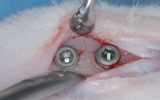
Fig. 2 Two implants were placed on each tibia.
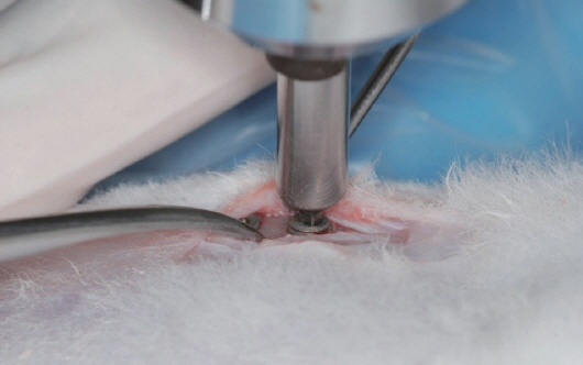
Fig. 3 Removal torque were measured by digital torque gauge.

Fig. 4 Comparison of 1st and 2nd removal torques in each group.

Fig. 5 Mean BIC in each group.
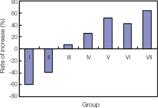
Fig. 6 Rate of increase between 1st and 2nd removal torques in each group.

Fig. 7 Histomorphometric analysis of control group and experimental groups. Tetracycline initially appeared on the bone surrounding the implant in Group II, and it became apparent in Group IIIand more as time passed (Green band; Green arrow). Demeclocycline continued to appear in Groups VI and VII (Yellow band; Yellow arrow).
Reference
-
References
1. Brånemark PI, Hansson BO, Adell R, Breine U, Lindström J, Hallén O, Ohman A. Osseointegrated implants in the treatment of edentulous jaw. Experience from a 10-years period. Scan J Plast Reconstr Surg Suppl. 1977; 16:1–132. PMID: 356184.2. Jang JH, Cho JH, Lee CH. Study of the re-osseointegration of implant fixture after mechanical unscrewing. J Korean Acad Prosthodont. 2010; 48:20914. DOI: 10.4047/jkap.2010.48.3.209.3. Hwang YJ, Cho JH, Lee CH. Investigation of osseointegration according to the healing time after having iatrogenic mobility of implant fixtures. J Korean Acad Prosthodont. 2010; 48:308–14. DOI: 10.4047/jkap.2010.48.4.308.4. Ivanoff CJ, Sennerby L, Lekholm U. Reintegration of mobilized titanium implants. An experimental study in rabbit tibia. Int J Oral Maxillofac Surg. 1997; 26:310–5. DOI: 10.1016/S0901-5027(97)80878-8.5. Cochran DL. A comparison of endosseous dental implant surfaces. J Periodontol. 1999; 70:1523–39. DOI: 10.1902/jop.1999.70.12.1523. PMID: 10632528.6. Sennerby L, Thomsen P, Ericson LE. A morphometric and biomechanic comparison of titanium implants inserted in rabbit cortical and cancellous bone. Int J Oral Maxillofac Implants. 1992; 7:62–71. PMID: 1398826.7. Roberts EW, Turley PK, Brezniak N, Fielder PJ. Implants: Bone physiology and metabolism. CDA J. 1987; 15:54–61. PMID: 3331974.8. Roberts EW. Bone tissue interface. Int J Oral Implantol. 1988; 5:71–4. PMID: 3271586.9. Roberts EW, Smith RK, Zilberman Y, Mozsary PG, Smith RS. Osseous adaptation to continuous loading of rigid endosseous implants. Am J Orthod. 1984; 86:95–111. DOI: 10.1016/0002-9416(84)90301-4.10. Sennerby L, Thomsen P, Ericson LE. Early tissue response to titanium implants inserted in rabbit cortical bone. Part I Light microscopic observations. J Mater Sci Mater Med. 1993; 4:240–50. DOI: 10.1007/BF00122275.11. Schwartz Z, Kieswetter K, Dean DD, Boyan BD. Underlying mechanisms at the bone-surface interface during regeneration. J Periodontal Res. 1997; 32:166–71. DOI: 10.1111/j.1600-0765.1997.tb01399.x. PMID: 9085228.
- Full Text Links
-
- Actions
-
Cited
- CITED
-
- Close
- Share
-
- Similar articles
-
- Investigation of osseointegration according to the healing time after having iatrogenic mobility of implant fixtures
- THREE DIMENSIONAL FINITE ELEMENT ANALYSIS ON THE MANDIBULAR CANTILEVERED PROSTHESIS SUPPORTED BY IMPLANTS
- Comparison of Complications in Direct and Indirect Osseointegration of Prosthetic Auricular Reconstruction
- Monitoring osseointegrated prosthesis loosening and fracture using electrical capacitance tomography
- Preliminary Study on the Design of Implant Abutment Screw Head for Rapid Fastening & loosening
