J Adv Prosthodont.
2014 Feb;6(1):53-59. 10.4047/jap.2014.6.1.53.
Residual bone height measured by panoramic radiography in older edentulous Korean patients
- Affiliations
-
- 1Department of Prosthodontics, College of Dentistry, Dankook University, Cheonan, Republic of Korea. cho8511@dankook.ac.kr
- KMID: 2176571
- DOI: http://doi.org/10.4047/jap.2014.6.1.53
Abstract
- PURPOSE
The aim of this study was to obtain statistical data on the residual bone height at different natural tooth positions by panoramic radiography in edentulous Korean patients aged 60-90 years.
MATERIALS AND METHODS
The study included the diagnostic panoramic radiographs of 180 randomly selected edentulous patients without systemic diseases affecting bone. The radiographic selection criteria included absence of obvious facial asymmetry, clearly visible anatomic structures, and no surgical and fracture history. The panoramic radiographs of 79 patients met these criteria and were used in the analysis. The same researcher processed all the radiographs by using a standardized method. The height of the residual bone was measured at 18 predetermined sites (7 in the maxilla and 11 in the mandible) on digitized and printed radiographs by using a Digimatic caliper, triangle, and ruler. Gender- and age-related differences were statistically analyzed by using the t-test and rank-sum test (alpha=0.05).
RESULTS
The maxillary residual bone height did not show significant gender-related differences, but male patients had significantly higher residual bone in the mandible(P<.05). No significant height differences at the measured sites were noted among the 60s, 70s, and 80s age groups.
CONCLUSION
Dentists should pay greater attention to older female edentulous patients because they are more prone to rapid residual bone resorption. Residual bone resorption may not be affected by age.
Keyword
MeSH Terms
Figure
-
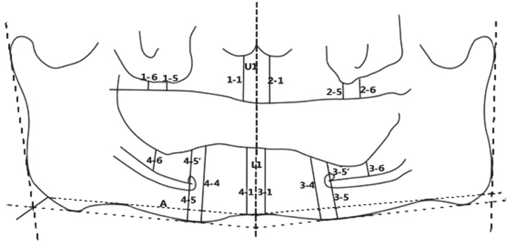
Fig. 1 Vertical distances used to measure the residual bone height of the edentulous patients.
Cited by 1 articles
-
Alveolar bone height according to the anatomical relationship between the maxillary molar and sinus
Yoon Joo Choi, Young Hyun Kim, Sang-Sun Han, Ui-Won Jung, Chena Lee, Ari Lee, Kug Jin Jeon
J Periodontal Implant Sci. 2020;50(1):38-47. doi: 10.5051/jpis.2020.50.1.38.
Reference
-
1. Tallgren A. The continuing reduction of the residual alveolar ridges in complete denture wearers: a mixed-longitudinal study covering 25 years. J Prosthet Dent. 1972; 27:120–132.2. Misch CE. Contemporary implant dentistry. 2nd ed. St. Louis: CV Mosby;1997. p. 3–12.3. Brodeur JM, Laurin D, Vallee R, Lachapelle D. Nutrient intake and gastrointestinal disorders related to masticatory performance in the edentulous elderly. J Prosthet Dent. 1993; 70:468–473.4. Soikkonen K, Ainamo A, Xie Q. Height of the residual ridge and radiographic appearance of bony structure in the jaws of clinically edentulous elderly people. J Oral Rehabil. 1996; 23:470–475.5. Lee SM, Lee SS, Huh KH, Yi WJ, Heo MS, Choi SC. The effects of location of alveolar crest on the vertical bone heights on panoramic radiographs. Dentomaxillofac Radiol. 2012; 41:117–121.6. Shin JW. Dental anatomy. 3rd ed. Seoul: DaehanNarae pub.;2010. p. 65–221.7. Thorpe JO. Panoramic radiography in the general practice of dentistry. Oral Surg Oral Med Oral Pathol. 1967; 24:781–792.8. Crane GM, Ishaug SL, Mikos AG. Bone tissue engineering. Nat Med. 1995; 1:1322–1324.9. Rowe DJ. Bone loss in the elderly. J Prosthet Dent. 1983; 50:607–610.10. Hirai T, Ishijima T, Hashikawa Y, Yajima T. Osteoporosis and reduction of residual ridge in edentulous patients. J Prosthet Dent. 1993; 69:49–56.11. Zarb GA, Bolender CL. Prosthodontic treatment for edentulous patients. 2nd ed. St.Louis: CV Mosby;1997. p. 10.12. Misch CE. Contemporary implant dentistry. 3rd ed. St. Louis: CV Mosby;2008. p. 939–951.13. Mahon JM, Norling BK, Phoenix RD. Effect of varying fixture width on stress and strain distribution associated with an implant stack system. Implant Dent. 2000; 9:310–320.14. Friberg B, Gröndahl K, Lekholm U, Brånemark PI. Long-term follow-up of severely atrophic edentulous mandibles reconstructed with short Brånemark implants. Clin Implant Dent Relat Res. 2000; 2:184–189.15. Aparicio C, Perales P, Rangert B. Tilted implants as an alternative to maxillary sinus grafting: a clinical, radiologic, and periotest study. Clin Implant Dent Relat Res. 2001; 3:39–49.16. Güler AU, Sumer M, Sumer P, Biçer I. The evaluation of vertical heights of maxillary and mandibular bones and the location of anatomic landmarks in panoramic radiographs of edentulous patients for implant dentistry. J Oral Rehabil. 2005; 32:741–746.17. Feine JS, Carlsson GE, Awad MA, Chehade A, Duncan WJ, Gizani S, Head T, Lund JP, MacEntee M, Mericske-Stern R, Mojon P, Morais J, Naert I, Payne AG, Penrod J, Stoker GT Jr, Tawse-Smith A, Taylor TD, Thomason JM, Thomson WM, Wismeijer D. The McGill Consensus Statement on Overdentures. Montreal, Quebec, Canada. May 24-25, 2002. Int J Prosthodont. 2002; 15:413–414.18. Devlin H, Ferguson MW. Alveolar ridge resorption and mandibular atrophy. A review of the role of local and systemic factors. Br Dent J. 1991; 170:101–104.19. Desjardins RP. Tissue-integrated prostheses for the edentulous patients: Branemark (Nobelpharma) System. In : Caswell CW, Clark AE, editors. Dental implant prosthodontics. Philadelphia: J.B. Lippincott Co;1991. p. 1–57.20. Mercier P. Ridge reconstruction with hydroxylapatite. Part 1. Anatomy of the residual ridge. Oral Surg Oral Med Oral Pathol. 1988; 65:505–510.
- Full Text Links
-
- Actions
-
Cited
- CITED
-
- Close
- Share
-
- Similar articles
-
- Bone height measurements of implant sites: Comparison of panoramic radiography and spiral computed tomography
- A new bite block for panoramic radiographs of anterior edentulous patients: A technical report
- Comparison of the reproducibility of panoramic radiographs between dentulous and edentulous patients
- Bone density relationship of mandible and cervical vertebrae in panoramic radiography
- Evaluation of dental panoramic radiographic findings in edentulous jaws: A retrospective study of 743 patients "Radiographic features in edentulous jaws"
