Comparison of the reproducibility of panoramic radiographs between dentulous and edentulous patients
- Affiliations
-
- 1Department of Oral and Maxillofacial Radiology and Dental Research Institute, School of Dentistry, Seoul National University, Seoul, Korea. hmslsh@snu.ac.kr
- KMID: 1799590
- DOI: http://doi.org/10.5624/isd.2014.44.2.95
Abstract
- PURPOSE
This study was performed to evaluate the reproducibility of panoramic radiographs of dentulous and edentulous patients.
MATERIALS AND METHODS
The reproducibility of panoramic radiographs was evaluated using the panoramic radiographs acquired from 30 anterior dentulous patients by using a common biting positioning device (dentulous group) and 30 anterior edentulous patients by using chin-support devices to take a panoramic radiograph (edentulous group), respectively; these patients had undergone 3 or more panoramic radiographs. The widths and angles between the designated landmarks were measured on the panoramic radiographs, and the reproducibility was evaluated using the intraclass correlation coefficient (ICC) and the coefficient of variation.
RESULTS
In the dentulous and edentulous groups, the ICCs of the mandibular ramus and mandibular angle areas were higher than the condylar head and zygomatic areas. The mandibular ramus and angle areas showed statistically lower mean coefficients of variation than the condylar head and zygomatic areas in the dentulous group. The mandibular angle area showed a significantly lower mean coefficient of variation than the zygomatic area in the edentulous group. By comparing the two groups, each ICC of the edentulous group was lower than that of the dentulous group, and the mean coefficients of variation of the mandibular ramus area, zygomatic area, left condylar inclination, and ramus ratio between the right and the left in the edentulous group were significantly higher than those in the dentulous group.
CONCLUSION
Biting positioning for dentulous patients provided better positioning reproducibility than chin-support positioning when performing panoramic radiography for edentulous patients.
Figure
-

Fig. 1 A. The conventional standard bite block is used for anterior dentulous patients. B. The conventional chin support-device is used for anterior edentulous patients.
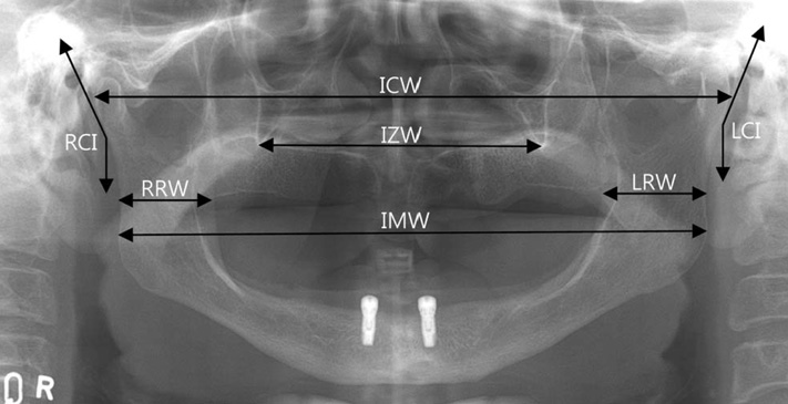
Fig. 2 Reference points, lines, and angles of measurement variables are seen on a panoramic radiograph. IMW: Width between the farthest distal mandibular angle points, ICW: Width between the farthest distal condyle head points, IZW: Width between the bottom points of the innominate line, RRW: Shortest horizontal length of the right mandibular ramus, LRW: Shortest horizontal length of the left mandibular ramus, RCI: Angle between a parallel line with the distal right condyle neck and a vertical line, LCI: Angle between a linear line with the distal left condyle neck and a vertical line.

Fig. 3 Intraclass correlation coefficients of widths and angles to show reliability on reproduced panoramic radiographs in the dentulous group and the edentulous group (The higher, the better).
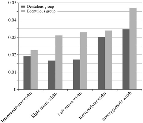
Fig. 4 Mean coefficient of variation of widths to show precision on reproduced panoramic radiographs in the dentulous and the edentulous groups. All the areas (Intermandibular width, Right ramus width, Left ramus width, Intercondylar width, and Interzygomatic width) of the dentulous group showed lower mean coefficients of variation, which indicate higher precision, than those of the edentulous group (The lower, the better).
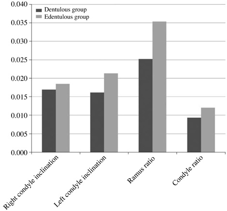
Fig. 5 Mean coefficient of variation of right condyle inclination, left condyle inclination, ratio between the right ramus width and the left ramus width (ramus ratio), and ratio between the right condyle inclination and the left condyle inclination (condyle ratio) on the reproduced panoramic radiographs in the dentulous and the edentulous groups, which shows the precision of the condyle inclination, and the ratio between the right and the left side or the precision of rotation on the vertical axis (The lower, the better).
Cited by 1 articles
-
A new bite block for panoramic radiographs of anterior edentulous patients: A technical report
Jong-Woong Park, Khanthaly Symkhampha, Kyung-Hoe Huh, Won-Jin Yi, Min-Suk Heo, Sam-Sun Lee, Soon-Chul Choi
Imaging Sci Dent. 2015;45(2):117-122. doi: 10.5624/isd.2015.45.2.117.
Reference
-
1. Kogon S, Bohay R, Stephens R. A survey of the radiographic practices of general dentists for edentulous patients. Oral Surg Oral Med Oral Pathol Oral Radiol Endod. 1995; 80:365–368.
Article2. Spyropoulos ND, Patsakas AJ, Angelopoulos AP. Findings from radiographs of the jaws of edentulous patients. Oral Surg Oral Med Oral Pathol. 1981; 52:455–459.
Article3. Swenson HM, Hudson JR. Roentgenographic examination of edentulous patients. J Prosthet Dent. 1967; 18:304–307.
Article4. Ortman LF, Hausmann E, Dunford RG. Skeletal osteopenia and residual ridge resorption. J Prosthet Dent. 1989; 61:321–325.
Article5. Soikkonen K, Ainamo A, Xie Q. Height of the residual ridge and radiographic appearance of bony structure in the jaws of clinically edentulous elderly people. J Oral Rehabil. 1996; 23:470–475.
Article6. Wical KE, Swoope CC. Studies of residual ridge resorption. I. Use of panoramic radiographs for evaluation and classification of mandibular resorption. J Prosthet Dent. 1974; 32:7–12.7. Kaffe I, Ardekian L, Gelerenter I, Taicher S. Location of the mandibular foramen in panoramic radiographs. Oral Surg Oral Med Oral Pathol. 1994; 78:662–669.
Article8. Choi BR, Choi DH, Huh KH, Yi WJ, Heo MS, Choi SC, et al. Clinical image quality evaluation for panoramic radiography in Korean dental clinics. Imaging Sci Dent. 2012; 42:183–190.
Article9. Welander U. A mathematical model of narrow beam rotation methods. Acta Radiol Diagn (Stockh). 1974; 15:305–317.
Article10. Millar JK. Dental pantomography. The orthopantomograph: a method of patient positioning. Radiography. 1979; 45:197–199.11. Tronje G, Welander U, McDavid WD, Morris CR. Image distortion in rotational panoramic radiography. I. General considerations. Acta Radiol Diagn (Stockh). 1981; 22:295–299.12. Tronje G, Eliasson S, Julin P, Welander U. Image distortion in rotational panoramic radiography. II. Vertical distances. Acta Radiol Diagn (Stockh). 1981; 22:449–455.13. Tronje G, Welander U, McDavid WD, Morris CR. Image distortion in rotational panoramic radiography. III. Inclined objects. Acta Radiol Diagn (Stockh). 1981; 22:585–592.14. Catić A, Celebić A, Valentić-Peruzović M, Catović A, Jerolimov V, Muretić I. Evaluation of the precision of dimensional measurements of the mandible on panoramic radiographs. Oral Surg Oral Med Oral Pathol Oral Radiol Endod. 1998; 86:242–248.15. Larheim TA, Svanaes DB, Johannessen S. Reproducibility of radiographs with the orthopantomograph 5: tooth-length assessment. Oral Surg Oral Med Oral Pathol. 1984; 58:736–741.
Article16. Larheim TA, Svanaes DB. Reproducibility of rotational panoramic radiography: mandibular linear dimensions and angles. Am J Orthod Dentofacial Orthop. 1986; 90:45–51.
Article17. Sämfors KA, Welander U. Angle distortion in narrow beam rotation radiography. Acta Radiol Diagn (Stockh). 1974; 15:570–576.
Article18. Zach GA, Langland OE, Sippy FH. The use of orthopantomograph in longitudinal studies. Angle Orthod. 1969; 39:42–50.19. Richardson JE, Langland OE, Sippy FH. A cephalostat for the orthopantomograph. Oral Surg Oral Med Oral Pathol. 1969; 27:643–646.
Article20. McDavid WD, Tronje G, Welander U, Morris CR. Effects of errors in film speed and beam alignment on the image layer in rotational panoramic radiography. Oral Surg Oral Med Oral Pathol. 1981; 52:561–564.
Article21. Scarfe WC, Eraso FE, Farman AG. Characteristics of the Orthopantomograph OP 100. Dentomaxillofac Radiol. 1998; 27:51–57.
Article22. Shrout PE, Fleiss JL. Intraclass correlations: uses in assessing rater reliability. Psychol Bull. 1979; 86:420–428.
Article23. Landis JR, Koch GG. The measurement of observer agreement for categorical data. Biometrics. 1977; 33:159–174.
Article24. Pfeiffer P, Bewersdorf S, Schmage P. The effect of changes in head position on enlargement of structures during panoramic radiography. Int J Oral Maxillofac Implants. 2012; 27:55–63.25. Hardy TC, Suri L, Stark P. Influence of patient head positioning on measured axial tooth inclination in panoramic radiography. J Orthod. 2009; 36:103–110.
Article
- Full Text Links
-
- Actions
-
Cited
- CITED
-
- Close
- Share
-
- Similar articles
-
- A new bite block for panoramic radiographs of anterior edentulous patients: A technical report
- Changes in Condylar Shape and Gonial Angle according to Loss of Teeth in Elderly Population
- Evaluation of median mandibular flexure values in dentulous and edentulous subjects by using an intraoral digital scanner
- Comparison of panoramic radiography and cone-beam computed tomography for assessing radiographic signs indicating root protrusion into the maxillary sinus
- Residual bone height measured by panoramic radiography in older edentulous Korean patients
