J Korean Acad Conserv Dent.
2007 Jul;32(4):385-396. 10.5395/JKACD.2007.32.4.385.
Comparison of shaping ability between single length technique and crown-down technique using Mtwo rotary file
- Affiliations
-
- 1Department of Conservative Dentistry, School of Dentistry, Pusan National University, Busan, Korea. golddent@pusan.ac.kr
- KMID: 2175900
- DOI: http://doi.org/10.5395/JKACD.2007.32.4.385
Abstract
- The aims of this study were to compare the shaping effect and safety between single length technique recommended by manufacturer and crown-down technique using Mtwo rotary file and to present a modified method in use of Mtwo file. Sixty simulated root canal resin blocks were used. The canals were divided into three groups according to instrument and the manner of using methods. Each group had 20 specimens. Group MT was instrumented with single length technique of Mtwo, group MC was instrumented with crowndown technique of Mtwo and group PT was instrumented with crown-down technique of ProTaper. All of the rotary files used in this study were operated by an electric motor. The scanned canal images of before and after preparation were superimposed. These superimposed images were evaluated at apical 1 to 8 mm levels. Angle changes were calculated. The preparation time, weight loss, instrument failure and binding, canal aberrations, and centering ratio were measured. Statistical analysis of the three experimental groups was performed with ANOVA and Duncan's multiple range tests for post-hoc comparison and Fisher's exact test was done for the frequency comparison. In total preparation time, group MT and group MC were less than group PT. In the aberrations, group MT had more elbows than those of group MC and group PT. The binding of group MC was least and group MT was less than group PT (P < 0.05). Under the condition of this study, crown-down technique using Mtwo rotary file is better and safer method than single length technique recommended by the manufacturer.
MeSH Terms
Figure
-

Figure 1 Purple; Pre-instrumented canal, Yellow; Post-instrumented canal Sample of superimposed image.

Figure 2 The representative images of three superimposed groups.
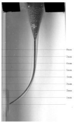
Figure 3 The horizontal lines mean the 8 measuring levels.
Cited by 1 articles
-
A comparison of the shaping ability of four rotary nickel-titanium files in simulated root canals
Bo-Hye Kim, Kyoung-Kyu Choi, Sang-Hyuk Park, Gi-Woon Choi
J Korean Acad Conserv Dent. 2010;35(2):88-95. doi: 10.5395/JKACD.2010.35.2.088.
Reference
-
1. Glossen CR, Haller RH, Dove SB, del Rio CE. A comparison of root canal preparations using Ni-Ti hand, Ni-Ti engine-driven, and K-Flex endodontic instruments. J Endod. 1995. 21:146–151.
Article2. Walia HM, Brantley WA, Gerstein H. An initial investigation of the bending and torsional properties of Nitinol root canal files. J Endod. 1988. 14:346–351.
Article3. Schäfer E, Schulz-Bongert U, Tulus G. Comparison of hand stainless steel and nickel titanium rotary instrumentation: a clinical study. J Endod. 2004. 30:432–435.
Article4. Chen JL, Messer HH. A comparison of stainless steel hand and rotary nickel-titanium instrumentation using a silicone impression technique. Aust Dent J. 2002. 47:12–20.
Article5. Garip Y, Gunday M. The use of computed tomography when comparing nickel-titanium and stainless steel files during preparation of simulated curved canals. Int Endod J. 2001. 34:452–457.
Article6. Pettiette MT, Delano EO, Trope M. Evaluation of success rate of endodontic treatment performed by students with stainless-steel K-files and nickel-titanium hand files. J Endod. 2001. 27:124–127.
Article7. Schäfer E. Shaping ability of Hero 642 rotary nickel-titanium instruments and stainless steel hand K-Flexofiles in simulated curved root canals. Oral Surg Oral Med Oral Pathol Oral Radiol Endod. 2001. 92:215–220.
Article8. Peters OA. Current Challenges and Concepts in the Preparation of Root Canal Systems: A Review. J Endod. 2004. 30:559–567.
Article9. Kum KY, Spängberg L, Cha BY, Jung IY, Lee SJ, Lee CY. Shaping ability of three ProFile rotary instrumentation techniques in simulated resin root canals. J Endod. 2000. 26:719–723.
Article10. Schäfer E, Erler M, Dammaschke T. Comparative study on the shaping ability and cleaning efficiency of rotary Mtwo instruments. Part 1. Shaping ability in simulated curved canals. Int Endod J. 2006. 39:196–202.
Article11. Veltri M, Mollo A, Mantovani L, Pini P, Balleri P, Grandini S. A comparative study of Endoflare-Hero Shaper and Mtwo NiTi instruments in the preparation of curved root canals. Int Endod J. 2005. 38:610–616.
Article12. Schneider SW. A comparison of canal preparations in straight and curved root canals. Oral Surg. 1971. 32:271–275.
Article13. Gunday M, Sazak H, Garip Y. A comparative study of three different root canal curvature measurement techniques and measuring the canal access angle in curved canals. J Endod. 2005. 31:796–798.
Article14. Kim HC, Park JK, Hur B. Relative efficacy of three Ni-Ti file systems used by undergraduates. J Korean Acad Conserv Dent. 2005. 30:38–48.
Article15. Bergmans L, Van Cleynenbreugel J, Beullens M, Wevers M, Van Meerbeek B, Lambrechts P. Progressive versus constant tapered shaft design using NiTi rotary instruments. Int Endod J. 2003. 36:288–295.
Article16. Calhoun G, Montgomery S. The effects of four instrumentation techniques on root canal shape. J Endod. 1988. 14:273–277.
Article17. Kosa DA, Marshall G, Baumgartner JC. An analysis of canal centering using mechanical instrumentation techniques. J Endod. 1999. 25:441–445.
Article18. Foschi F, Nucci C, Montebugnoli L, Marchionni S, Breschi L, Malagnino VA, Prati C. SEM evaluation of canal wall dentine following use of Mtwo and ProTaper NiTi rotary instruments. Int Endod J. 2004. 37:832–839.
Article19. Schäfer E, Erler M, Dammaschke T. Comparative study on the shaping ability and cleaning efficiency of rotary Mtwo instruments. Part 2. Cleaning effectiveness and shaping ability in severely curved root canals of extracted teeth. Int Endod J. 2006. 39:203–212.
Article20. Berutti E, Chiandussi G, Gaviqlio I, Ibba A. Comparative analysis of torsional and bending stresses in two mathematical models of nickel-titanium rotary instruments: ProTaper versus ProFile. J Endod. 2003. 29:15–19.
Article21. Calberson FL, Deroose CA, Hommez GM, De Moor RJ. Shaping ability of ProTaper nickel-titanium files in simulated resin root canals. Int Endod J. 2004. 37:613–623.
Article22. Iqbal MK, Firic S, Tulcan J, Karabucak B, Kim S. Comparison of apical transportation between ProFile and ProTaper NiTi rotary instruments. Int Endod J. 2004. 37:359–364.
Article23. Schäfer E, Vlassis M. Comparative investigation of two rotary nickel-titanium instruments: ProTaper versus RaCe. Part 1. Shaping ability in simulated curved canals. Int Endod J. 2004. 37:229–238.
Article24. Yun HH, Kim SK. A comparison of the shaping abilities of 4 nickel-titanium rotary instruments in simulated root canals. Oral Surg Oral Med Oral Pathol Oral Radiol Endod. 2003. 95:228–233.
Article25. Peters OA, Peters CI, Schöneberger K, Barbakov F. ProTaper rotary root canal preparation: effect of canal anatomy on final shape analyzed by micro CT. Int Endod J. 2003. 36:86–92.
Article26. Hong ES, Park JK, Hur B, Kim HC. Comparison of shaping ability between various hybrid instrumentation methods with ProTaper. J Korean Acad Conserv Dent. 2006. 31:11–19.
Article27. Peters OA, Peters CI, Schönenberger K, Barbakow F. assessment of torque and force in relation to canal anatomy. Int Endod J. 2003. 36:93–99.28. Diemer F, Calas P. Effect of pitch length on the behaviour of rotary triple helix root canal instruments. J Endod. 2004. 30:716–718.
Article29. Bergmans L, Van Cleynenbreugel J, Wevers M, Lambrechts P. Mechanical root canal preparation with NiTi rotary instruments: Rationale, performance and safety. Status report for the American Journal of Dentistry. Am J Dent. 2001. 14:324–333.30. Leeb JI. Canal orifice enlargement as related to biomechanical preparation. J Endod. 1983. 9:463–470.
Article31. Schrader C, Peters OA. Analysis of torque and force with differently tapered rotary endodontic instruments in vitro. J Endod. 2005. 31:120–123.
Article32. Sattapan B, Nervo GJ, Palamara JE, Messer HH. Defects in rotary nickel-titanium files after clinical use. J Endod. 2000. 26:161–165.
Article33. Cohen S, Burns RC. Pathways of the pulp. 1999. 7th edition. Translation Shinhung International, Inc.;252–254.
- Full Text Links
-
- Actions
-
Cited
- CITED
-
- Close
- Share
-
- Similar articles
-
- A comparison of the shaping ability of four rotary nickel-titanium files in simulated root canals
- Comparison of canal shaping methods with GT(TM) rotary file and condensation methods
- A comparative study of the canal configuration after shaping by protaper rotary and hand files in resin simulated canals
- Shaping ability of four rotary nickel-titanium instruments to prepare root canal at danger zone
- Comparison of shaping ability using various Nickel-Titanium rotary files and hybrid technique
